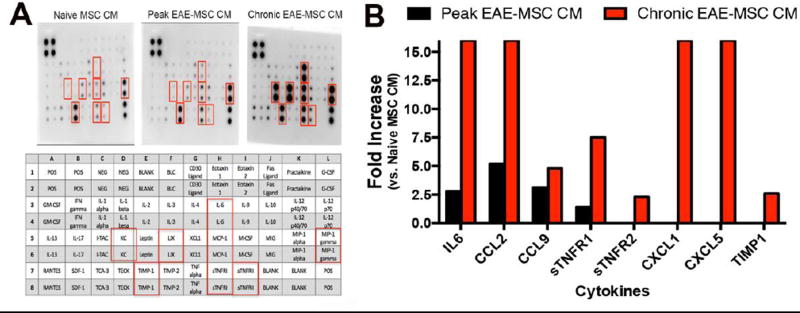
Figure 3. EAE-MSCs secrete higher levels of pro-inflammatory cytokines.
(A) Representative images of antibody arrays treated with conditioned medium (CM) from naïve MSCs, peak EAE-MSCs, or chronic EAE-MSCs. Cytokines found to be up-regulated in the arrays are indicated by red boxes and identified in the array diagram depicted below; CCL2 = MCP-1, CCL9 = MIP-1 gamma, CXCL1 = KC, and CXCL5 = LIX. (B) Fold changes of proteins increased in conditioned medium from peak or chronic EAE-MSCs relative to naïve MSCs; note conditioned medium from EAE-MSCs contains higher levels of pro-inflammatory cytokines, including IL6, CCL2, and CCL9. Fold changes were calculated by densitometric quantification of spot intensity values from 3 separate antibody arrays per group, n = 3 experiments. Fold changes were capped at 16, with no fold decreases of any cytokines tested observed in the arrays.

