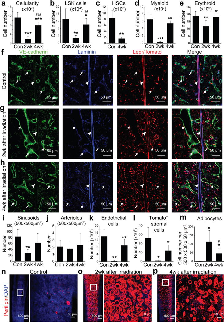
Figure 1. Irradiation disrupted sinusoids and depleted HSCs, endothelial cells, and LepR+ stromal cells while dramatically increasing adipocytes in the bone marrow.

One million bone marrow cells from wild-type mice were transplanted into irradiated wild-type (a–e and m–p) or Leprcre; R26tdTomato (f–l) mice. Statistical significance was assessed using repeated measures one-way ANOVAs with Geisser-Greenhouse sphericity corrections along with Tukey’s multiple comparisons tests (a–e, i–m). * indicates statistical significance relative to control (Con) while # indicates statistical significance of differences between 2 and 4 weeks after irradiation (* or # P<0.05, ** or ## P<0.01, *** or ### P<0.001). All data represent mean±SD.
(a–e) Flow cytometric analysis of mechanically dissociated bone marrow cells revealed significant reductions in bone marrow cellularity (a) and the numbers of Lineage−Sca-1+c-kit+ (LSK) cells (b), CD150+CD48−Lineage−Sca-1+c-kit+ HSCs (c), Mac1+Gr-1+ myeloid cells (d) and Ter119+ erythroid cells (e) at 2 and/or 4 weeks after irradiation as compared to non-irradiated control mice. Cell numbers reflect two femurs and two tibias per mouse (n=5 mice/treatment from 5 independent experiments).
(f–h) Confocal imaging of thin femur sections from non-irradiated Leprcre; R26tdTomato mice (control, f) or at 2 weeks (g) or 4 weeks (h) after irradiation and bone marrow transplantation. Arrows indicate sinusoidal blood vessels and arrowheads indicate arterioles.
(i, j) The densities of VE-cadhernbrightlaminindim sinusoids (i) and VE-cadherindimlamininbright arterioles (j) were quantified in sections (n=5 mice/condition from 3 independent experiments).
(k, l) Flow cytometric analysis of enzymatically dissociated bone marrow cells from Leprcre; R26tdTomato mice revealed significant reductions in the numbers of VE-cadherin+ endothelial cells (k) and Tomato+ stromal cells (l) after irradiation (n=4 mice/condition from 4 independent experiments).
(m) Adipocyte numbers in thick femur sections from non-irradiated mice (Con) or mice at 2 or 4 weeks after irradiation (n=6 mice/condition from 3 independent experiments).
(n–p) Whole-mount imaging of thick femur sections (50-μm) from non-irradiated mice (Control, n) or mice 2 (o) or 4 (p) weeks after irradiation and bone marrow transplantation. Adipocytes were identified based on anti-perilipin staining (n=6 mice/condition from 3 independent experiments).
