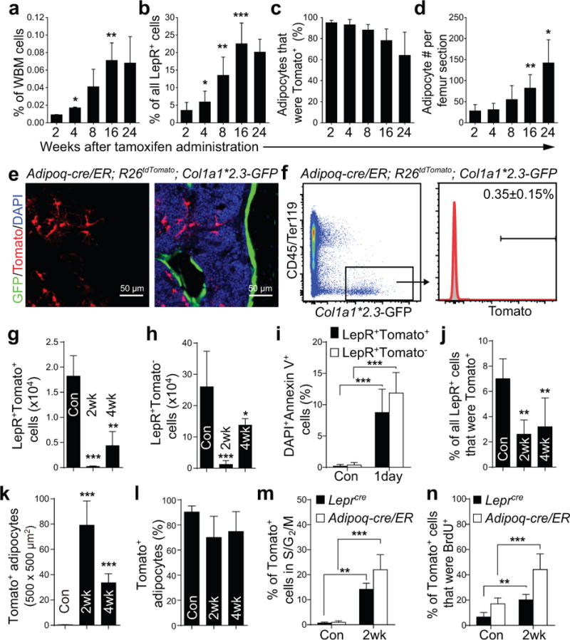
Figure 5. Adipoq-Cre/ER+ bone marrow stromal cells form adipocytes, but rarely osteoblasts, in vivo.

(a–d) Percentages of whole bone marrow (WBM) cells (a; by flow cytometry), LepR+ stromal cells (b; by flow cytometry), or adipocytes (c; by microscopy in sections) that were Tomato+ in Adipoq-cre/ER; R26tdTomato mice at 2–24 weeks after tamoxifen administration. Numbers of adipocytes/section (d). Two-way ANOVAs with Sidak’s multiple comparisons tests were used to assess differences among consecutive ages (*P<0.05, **P<0.01, ***P<0.001).
(e, f) Four months after tamoxifen treatment, Adipoq-cre/ER; R26tdTomato; Col1a1*2.3-GFP mice had only rare GFP+ osteoblasts that were Tomato+ in bone marrow sections (e) or by flow cytometry (f).
(g, h) Based on flow cytometric analysis, the numbers of LepR+Tomato+ cells (g) and LepR+Tomato− cells (h) declined in Adipoq-cre/ER; R26tdTomato mice after irradiation and bone marrow transplantation. Differences between non-irradiated (Con) and irradiated mice (at 2 or 4 weeks) were assessed by one-way ANOVAs with Dunnett’s multiple comparisons tests (data from panel g were log2-transformed) (**p<0.01, ***P<0.001).
(i) Percentages of LepR+Tomato+ or LepR+Tomato− cells that were also DAPI+Annexin V+ in enzymatically dissociated bone marrow cells from Adipoq-cre/ER; R26tdTomato mice one day after irradiation. A two-way ANOVA with Sidak’s multiple comparisons test was used to assess differences among treatments.
(j, k) The percentages of LepR+ cells that were Tomato+ (j) declined in Adipoq-cre/ER; R26tdTomato mice after irradiation. In contrast, the number of Tomato+ adipocytes in femur sections increased after irradiation (k). The statistical significance of differences between non-irradiated (Con) and irradiated mice was measured using one-way ANOVAs with Dunnett’s multiple comparisons tests (data of 5k were log2-transformed) (**P<0.01, ***P<0.001).
(l) The vast majority of bone marrow adipocytes were Tomato+ in non-irradiated and irradiated Adipoq-cre/ER; R26tdTomato mice.
(m, n) Hoechst staining (m) and BrdU incorporation (14 day pulse, n) by Tomato+ bone marrow stromal cells in Adipoq-cre/ER; R26tdTomato mice or Leprcre; R26tdTomato mice that were non-irradiated (Con) or at 2 weeks after irradiation and bone marrow transplantation. Two-way repeated measures ANOVAs with Sidak’s multiple comparisons tests was used to assess differences among treatments.
All data in Figure 5 represent mean±SD from n=5 mice/time point from 3 independent experiments.
