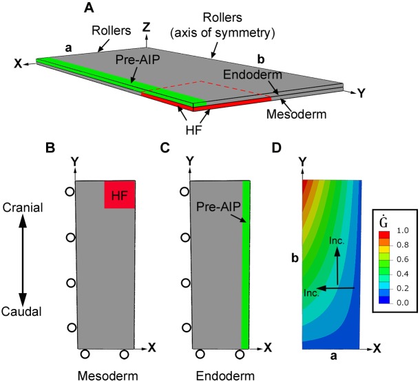
Fig. 5.
Finite-element model for foregut and heart tube formation in initial configuration (HH5). (A) The model for the inner splanchnopleure (iSPL) is a flat plate consisting of two layers representing the mesoderm and endoderm. Because of bilateral symmetry, only half of the iSPL is modeled with the y-axis representing the embryonic midline. Roller boundary conditions are indicated; other surfaces are free. (B,C) Ventral views of mesoderm and endoderm layers. Black circles indicate roller boundary conditions. Red and green denote regions representing the heart field (HF) and presumptive anterior intestinal portal (AIP), respectively. (D) The spatial pattern of growth rate ( , see Eqn S6) is the same for both layers during Phase 1, when diagonal folding of iSPL occurs. Growth rate is normalized to the maximum rate in each layer, but actual growth rates are higher in mesoderm (see supplementary Materials and Methods). Arrows indicate directions of increasing (Inc.) growth rate.

