Fig. 4.
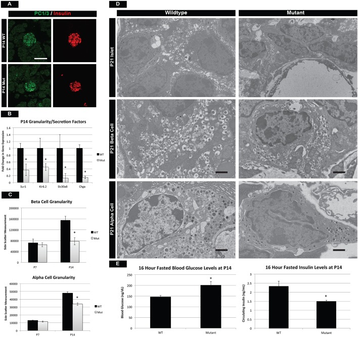
Mutant islet cells show reduced granularity and cellular function. (A) Prohormone convertase 1/3 (PC1/3) staining reveals proper insulin-processing machinery in β cells of mTOR mutants. Scale bar: 50 µm. (B) However, qPCR for endocrine transporters and granularity factors shows significant reduction in mRNA for the zinc transporter Slc30a8, which is involved in β-cell granule formation. This is also reflected in the decreased transcription of chromogranin A (Chga), a marker of intracellular vesicles. In addition, components (Sur1 and Kir6.2) of a K+ transporter necessary for insulin release are also downregulated in mutants. (C) Assessment of α- and β-cell granularity by average side-scatter measurements reveals normal granule formation at P7 but attenuated granularity at P14. (D) TEM at P21 confirms that mutant β and α cells have reduced numbers of insulin and glucagon granules, respectively. Scale bars: 1 µm. (E) These defects culminate in the inability of mutant β cells to respond to fasting glucose requirements at P14. See also Fig. S5. Mean±s.e.m.; *P<0.05. See Table S3 for numbers of animals used.

