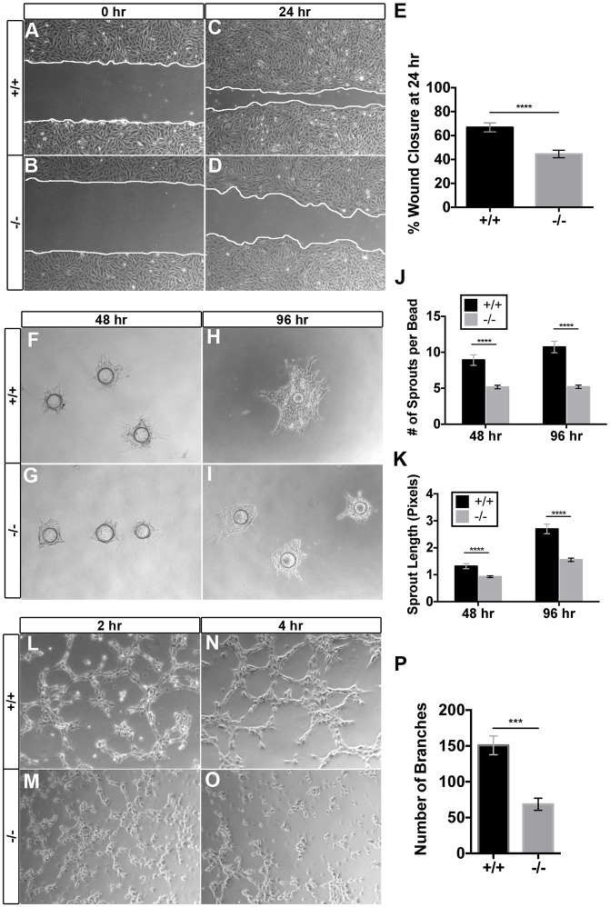
Fig. 5.
Egfl7−/− placental endothelial cells exhibit defects in cell migration, sprouting and branching morphogenesis. Isolated placental endothelial cells (ECs) from C57BL/6J (+/+) and Egfl7−/− mice were used for functional assays in vitro. (A-D) Representative images of the placental ECs from the scratch-wound assay. Collective EC migration was observed at 24 h (C,D). White lines indicate the leading edge of migrating cells. (E) Quantification of the percentage of wound area closed at 24 h (+/+, n=18; −/−, n=18). (F-I) Representative images of C57BL/6J (+/+) and Egfl7−/− placental ECs subjected to a bead sprouting assay, imaged at 48 h (F,G) and 96 h (H,I). (J,K) Quantification of the number of sprouts per bead (J) and the total sprout length (K) (+/+, n=11-12; −/−, n=20-23). (L-O) Representative images of C57BL/6J (+/+) and Egfl7−/− placental ECs subjected to a cord formation assay. Formation of cord-like structures on matrigel was observed at 2 h (L,M) and 4 h (N,O). (P) Quantification of the number of branches observed at 4 h, demonstrating a significant decrease in cord formation in Egfl7−/− placental ECs (+/+, n=20; −/−, n=15). Student's t-test; data are mean±s.e.m. ***P<0.001, ****P<0.0001. See also Figs. S2 and S4.

