Fig. 6.
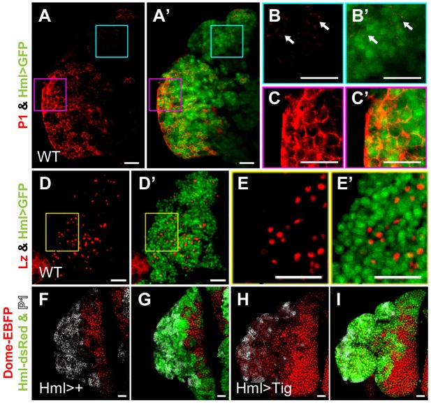
PLs contain a pool of Hml+/P1− IPs that is expanded by Tig expression. Confocal images of PLs from mid/late 3rd instar larvae. (A,A′) Stack projections of the surface layer of a P[Hml-Gal4]/P[UAS-GFP] PL with GFP (green) and P1 immunostaining (red). (B-C′) Magnified views illustrating areas with mostly IPs, i.e. GFP+ with no detectable P1 (B,B′; arrows indicate cells with low P1 levels) or largely mature plasmatocytes, i.e. GFP+ P1+ (C,C′). (D-E′) Stack projection of the surface layer of a wild-type PL expressing Hml>GFP (green) and immunostained for Lz (red). Crystal cells (Lz+) typically have little or no GFP, suggesting that the IPs are not crystal cells. Twelve PLs were examined for each condition. (F-I) PLs expressing P[Hml-Gal4] with or without P[UAS-Tig], also containing zone markers Hml-dsRed (green) and Dome-EBFP (red), and stained for P1 (white). Hml>Tig expanded the population of Hml+ P1− cells, which also contained intermediate levels of Dome-EBFP. See Table 2 for further quantification. There was also a reproducible increase in Dome-EBFP expression in the MZ (compare F with H), the reason for which is not clear. All animals were reared at 29°C. Scale bars: 25 µm.

