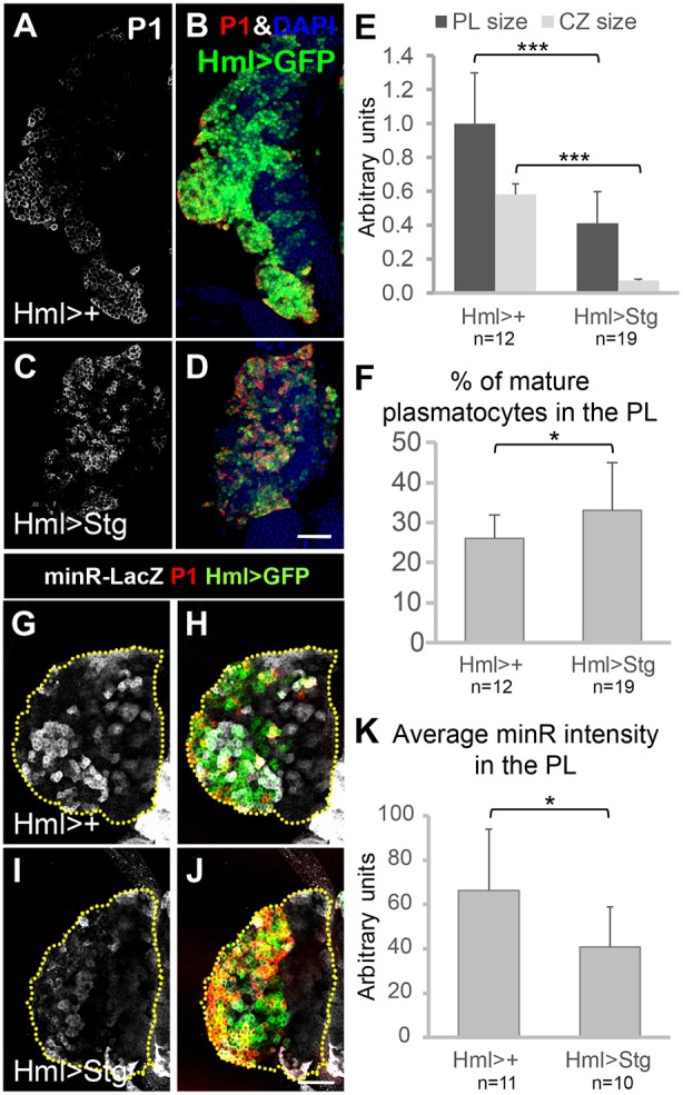
Fig. 9.

The phosphatase Stg promotes plasmatocyte differentiation and inhibits Tig reporter expression. (A-D) Confocal images of mid/late 3rd instar PLs containing P[Hml-Gal4] and P[UAS-GFP] with or without P[UAS-Stg], and immunostained for P1. In Hml>Stg PLs, the vast majority of Hml>GFP+ cells were also P1+, and the IP population (GFP+, low/no P1) was greatly reduced. (E) Quantification confirmed that PL and CZ sizes were greatly reduced in Hml>Stg PLs. (F) Quantification of P1 staining demonstrated an increase in mature plasmatocytes in Hml>Stg PLs. (G-J) Confocal images of mid 3rd instar PLs containing the minR-lacZ reporter, along with P[Hml-Gal4] and P[UAS-GFP] with or without P[UAS-Stg]. Stg expression inhibited minR-lacZ expression. (K) Quantification of the data in G-J. All animals were reared at 29°C. Data are mean±s.e.m. *P<0.05; ***P<0.001. Scale bars: 50 µm.
