Fig. 8.
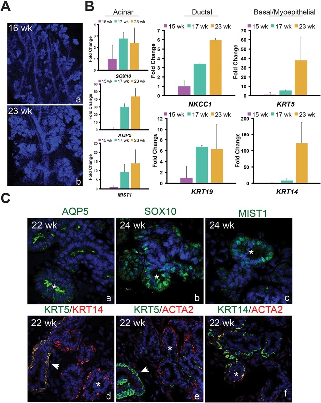
Phenotypic and molecular features of developing human and murine lacrimal glands are highly conserved. (A) The human fetal lacrimal gland undergoes extensive epithelial branching between 16 weeks (a) and 23 weeks (b). Scale bar: 500 μm. (B) qPCR analysis showed human lacrimal glands express several lineage markers observed in the murine lacrimal gland. All qPCR experiments used three biological replicates (data are mean±s.e.m.). (C) Immunostaining revealed that AQP5, SOX10 and MIST1 localize to the acini of fetal human lacrimal gland. As in mouse tissue, AQP5 was enriched in the apical membrane of acini and small ducts (a). SOX10 and MIST1 were exclusively found in acinar cells (b,c). Unlike in the murine lacrimal gland, KRT5 was barely detectable in myoepithelial cells when compared with basal cells, whereas KRT14 protein levels were similar in both cell types (d-f). Arrowheads indicate ducts and asterisks indicate acini.

