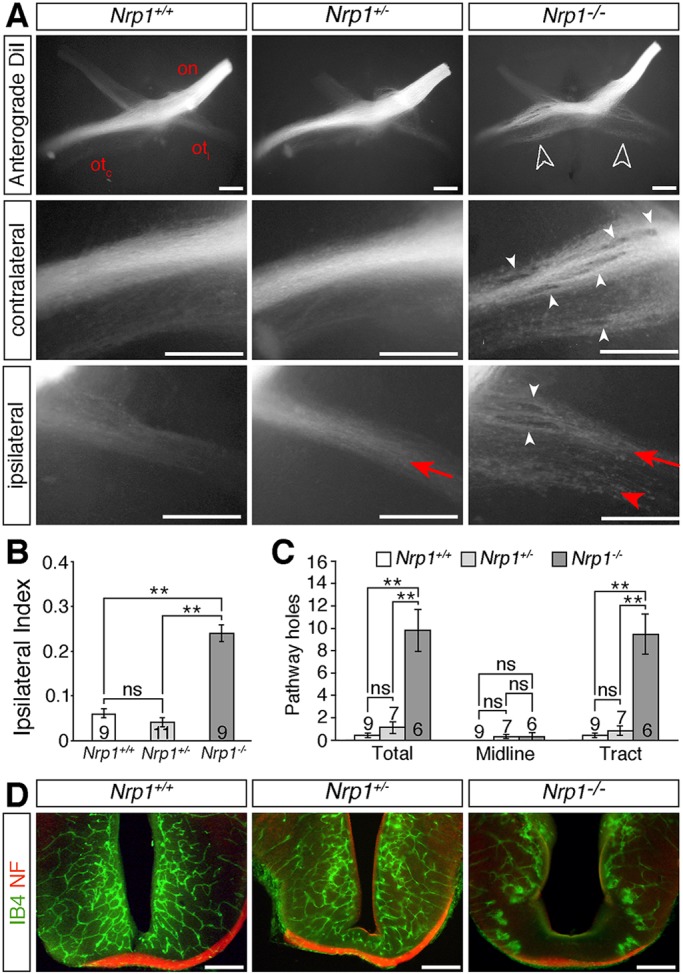
Fig. 1.

NRP1 is essential for normal development of the optic pathway. (A) Whole-mount view of RGC axons at the optic chiasm and contralateral and ipsilateral optic tracts in E14.5 Nrp1 wild-type (Nrp1+/+), heterozygous (Nrp1+/−) and null (Nrp1−/−) littermates, labelled anterogradely from one eye with DiI. Ventral view (top panels) or lateral view (middle and bottom panels), anterior up; on, optic nerve; otc, contralateral optic tract; oti, ipsilateral optic tract. White unfilled arrowheads indicate the increased proportion of ipsilaterally projecting RGC axons in mutants, white arrowheads indicate axonal exclusion zones, red arrows the normal position and organisation of the ipsilateral projection, red arrowhead ipsilateral optic tract defasciculation in mutants. (B,C) Ipsilateral index (B) and total number of exclusion zones (holes) at the optic chiasm (midline) and in the contralateral optic tract (C) in E14.5 Nrp1 wild-type, heterozygous and homozygous mutant littermates (mean±s.e.m.). **P<0.01; ns, not significant (one-way ANOVA with post-hoc Tukey). Numbers on bars indicate the number of embryos analysed for each genotype. (D) Coronal sections through the optic chiasm of E14.5 Nrp1 wild-type, heterozygous and homozygous mutant littermates labelled with isolectin B4 (IB4) to visualise blood vessels (green) and antibodies against neurofilaments (NF) to visualise nerves (red). Scale bars: 250 µm.
