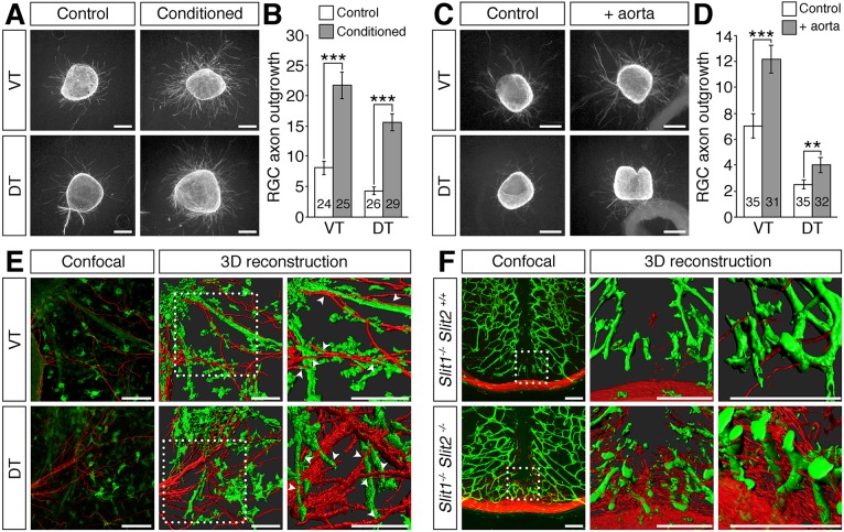
Fig. 8.
Vessels are not inhibitory to RGC axon growth in vitro or in vivo. (A-F) Retinal explants from E14.5 wild-type ventrotemporal (VT; source ipsilateral RGCs) or dorsotemporal (DT; source contralateral RGCs) retina cultured for 48 h in collagen gels with control or endothelial cell-conditioned medium (A,B) or co-cultured with an aortic ring (C,D). Cultures were stained for β-tubulin (A,C) and axon outgrowth quantified (B,D). Data are the mean±s.e.m. of at least three independent experiments. The number of explants analysed for each condition is indicated on each bar. ***P<0.001, **P<0.01 (Student's two-tailed unpaired t-test). In E, blood vessels were labelled with IB4 (green) and nerves with antibodies against neurofilaments (NF; red) and are shown as confocal z-stacks (left) and 3D reconstructions of the z-stacks (centre). The boxed regions are shown at higher magnification (right). Arrowheads indicate axons growing across or along the surface of vessels. (F) Confocal z-stacks and 3D reconstructions of the boxed region in the z-stacks at different magnifications of coronal sections through the diencephalon of E13.5 slit mutants. Blood vessels were visualised with IB4 (green) and nerves with antibodies against neurofilaments (red). Scale bars: 250 µm (A,C); 100 µm (E,F).

