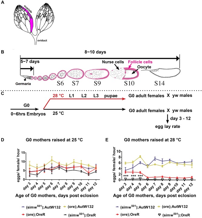
Fig. 1.
The (ore); OreR and (simw501); OreR females have a lower oviposition rate at a higher temperature. (A) An illustration of a pair of Drosophila ovaries with one ovariole indicated in pink. (B) A single ovariole with the developmental timeline of Drosophila oogenesis. Somatic follicle cells (pink) surround the germline nurse cells and oocyte to form an egg chamber. (C) Experimental scheme for the temperature-shift and female egg lay rate assay. (D,E) Oviposition rate of the indicated mito-nuclear females raised at 25°C (D) or 28°C (E) measured over 1 h. Fifty females per genotype, n=six biological replicates; data are mean±s.e.m. ***P<0.001 comparing (simw501); OreR with (ore); OreR using two-way ANOVA with Bonferroni correction. A and B are adapted, with the permission of the Genetics Society of America, from Ables (2015).

