Fig. 1.
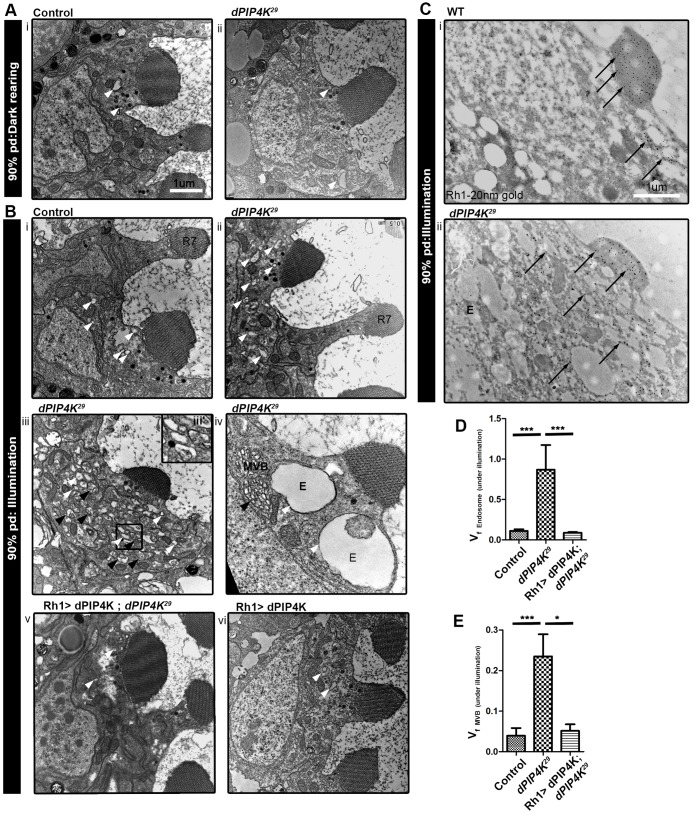
Loss of PIP4K leads to the formation of expanded endomembranes under illumination. (A,B) TEMs showing transverse sections of PRs from control (Ai) and PIP4K29 (Aii) pupae during late (90%) pupal development (pd) reared in the dark; and control (Bi), PIP4K29 (Bii–iv), Rh1>PIP4K; PIP4K29 (Bv) Rh1>PIP4K PRs (Bvi) reared under illumination. The white arrowheads indicate endosome-like vesicles (labelled E) and black arrowheads indicate the MVBs, both of which are expanded in PIP4K29 PRs under illumination (Bii–iv). R7, R7 PR cell. (C) Immuno-gold labelling for Rh1 in WT (i) and PIP4K29 PRs (ii). Arrows point to immunogold particles labelling Rh1, which are mostly present in the rhabdomere or apical plasma membrane (APM) in WT and in cytosolic endosome-like vesicles in PIP4K29 PRs. Scale bars: 1 μm. (D) Volume fraction analysis (VFA) for endosome-like vesicles in control, PIP4K29 and Rh1>PIP4K; PIP4K29 PRs under illumination at 90% pupal development. (E) VFA for MVBs under illumination at 90% pupal development. The values indicate the mean±s.e.m. of volume fraction in arbitrary units. *P<0.05; ***P<0.001 (one-way ANOVA with a nonparametric with Kruskal–Wallis test and Dunn's post test to compare the variance across samples).

