Fig. 4.
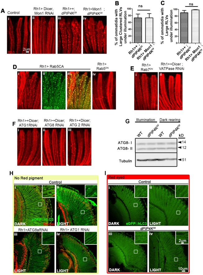
Unaltered endo-lysosomal flux in PIP4K29. (A) Rh1 immunostaining (red), in control (i) and Rh1>Mon1 RNAi lines (ii) showing an increase in the number of small RLVs with respect to the number seen in WT controls. RLVs stained in (iii) PIP4K29 and (iv) Rh1>Mon1; PIP4K29 are comparable. (B,C) Quantification of large clustered RLVs and large RLVs in Rh1>Mon1; PIP4K29 versus PIP4K29. (D) Rh1>YFP-Rab5CA (constitutively active) flies show an increase in the number of large RLVs (i–iii); these RLVs are not as expanded or clustered as is observed in the PIP4K29 line. (iv) There are fewer RLVs (red) in Rh1>Rab5DN than in WT. (E) (i) Rh1>Rab7DN shows an increase in the number of small RLVs with respect to WT. (ii) Rh1>V-ATPase (Vha44) RNAi causes no alteration in RLVs. (F) Downregulating components of the canonical autophagy pathway [Rh1>ATG1 (i), ATG8 (ii) and ATG2 (iii)] causes no change in RLV organization. (G) Western blot analysis of autophagy. Head extracts from PIP4K29 flies reared under illumination or in the dark showed no alteration in the autophagosome-associated, lipidated ATG8-II band with respect to that seen in WT reared under the same conditions. (H,I) Analysis of autophagy with the eGFP::hLC3 reporter. (H) Flies lacking the red (WT) screening pigment from the retinae, when exposed to constant illumination, showed accumulation of eGFP::hLC3 (green) in autophagosomes as punctae (ii and inset) whereas those reared in the dark did not, suggesting that autophagy is induced under illumination in the absence of screening pigment in the eye. ATG8 and ATG1 RNAi in the retinae under the similar conditions inhibit LC3 puncta formation (iii, iv). (I) WT flies (with the red screening pigment) showed no increase in eGFP::hLC3 puncta when reared under illumination compared to that seen upon dark rearing (ii versus i); PIP4K29 mutants, which also have an equivalent red eye colour (screening pigment), showed no change in LC3 puncta under illumination, implying no change in autophagic flux in red-eyed flies under light versus dark rearing. ns, not significant.

