
Figure 4. Analysis of VSELs by immunofluorescent staining and ImageStream System.
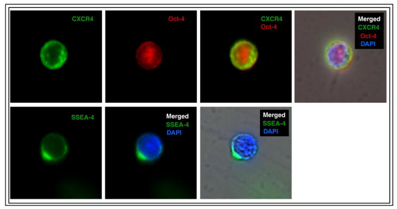
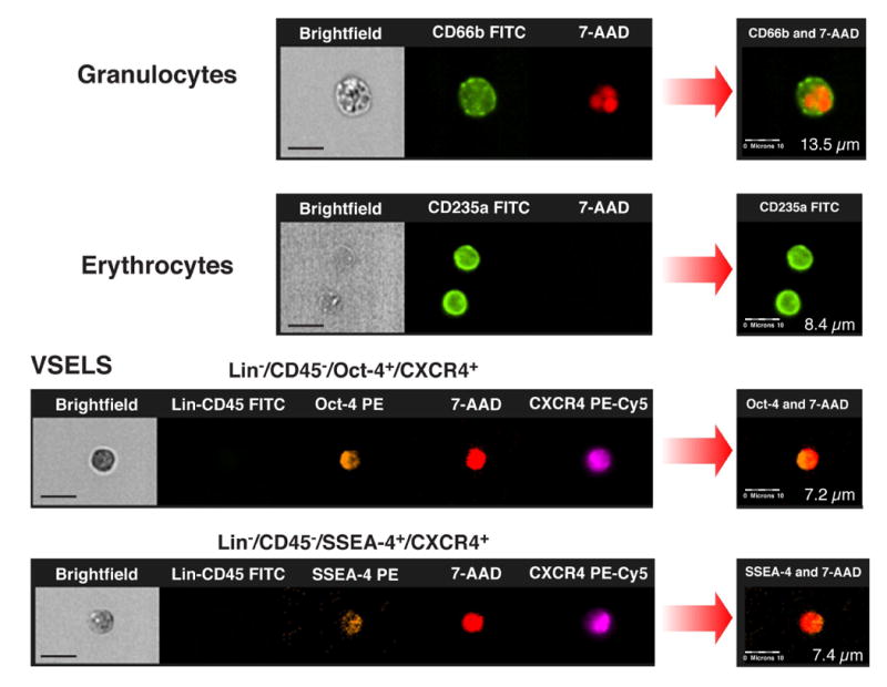
Immunofluorescent staining of peripheral blood-derived lin−CD45−CD133+ VSELs sorted with FACSAria (Panel A). VSELs express SSEA-4, Oct-4 and CXCR4 at the protein level. CXCR4 and SSEA-4 are visualized in the cellular membrane and Oct-4 in the nucleus. Nuclei are stained with DAPI. Staining was performed on cells isolated from four independent sorts. (Panel B) Circulating human VSELs analyzed by ImageStream system. Expression of Oct-4 and SSEA-4 in lin−CD45−CXCR4+ VSELs was confirmed at the single cell level. The size of VSELs was compared to granulocytes and erythrocytes derived from peripheral blood of patients with acute MI. The size of the cells was calculated by IDEAS software. Scale bars indicate 10μm.