
Fig. 3.
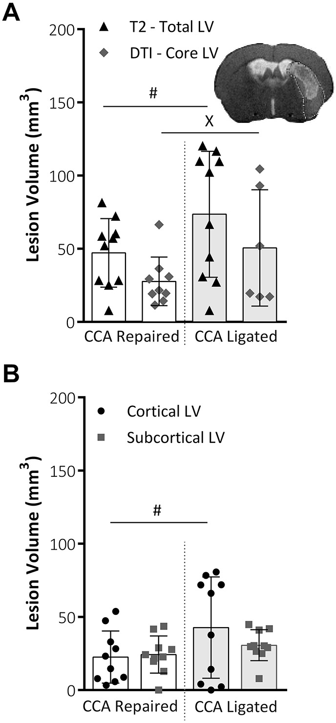
MRI lesion volume analysis. (A) Total lesion volume (mm3) at 48 h post-MCAO, as taken from T2-weighted MRI scans and core lesion volume as taken from DTI analysis (A). Variability within data, assessed using the F-test for parametric data or Lavene's test for non-parametric data, was significantly reduced for both total LV, i.e. T2-weighted scans, (P=0.015; CCA repair, n=10; CCA ligated, n=10; F-test) and core LV, i.e. DTI scans, (P=0.043; CCA repair, n=9; CCA ligated, n=6; Lavene's test). Representative image shows total lesion area from T2 scan slice with DTI core lesion volume mask overlaid. (B) Total lesion volume (mm3) at 48 h post-MCAO, taken from T2 MRI slices, split into cortical and subcortical lesion areas. CCA repair significantly reduced variability (P=0.03, F-test) in the cortical portion of lesion but did not affect variability in the sub-cortical portion of the lesion. Both groups, n=10. All data shown are mean±s.d., #P<0.05, F-test; XP<0.05, Lavene's test.