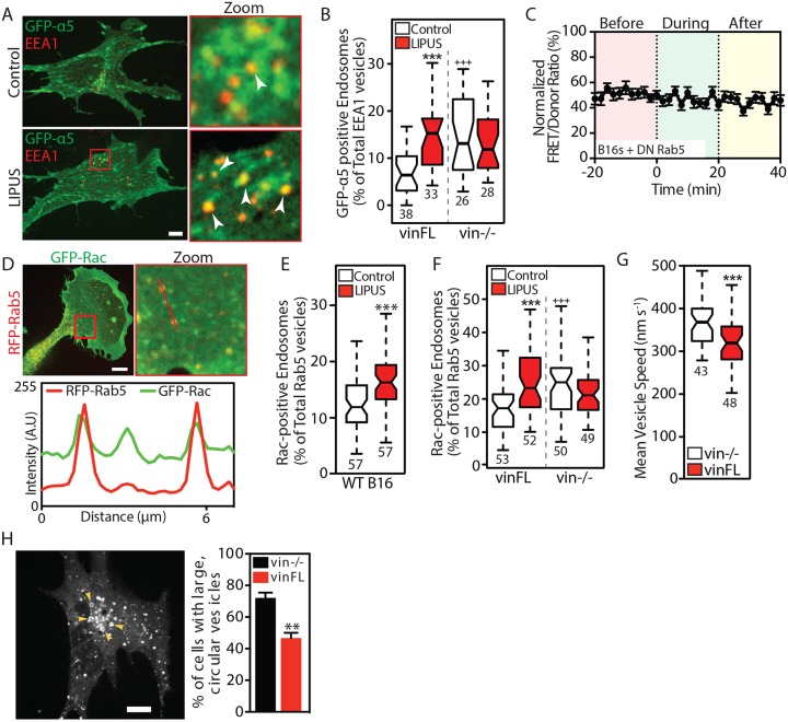
Fig. 7.
Vinculin modulates Rab5-mediated Rac1 activation in response to LIPUS. (A) Confocal images of vin−/– MEFs rescued with CFP-vinculin and coexpressing GFP-α5 integrin, without (top row) or with (bottom row) LIPUS stimulation, fixed and stained for EEA1. Arrowheads indicate colocalization between GFP-α5 and EEA1. Scale bar: 10 µm. (B) Colocalization between GFP-α5 and EEA1 in cells fixed 10 min after the end of LIPUS stimulation. ***P<0.001 against the control; +++P<0.001 against the vinFL control. (C) LIPUS stimulation has no effect on Rac1 activity in B16 cells expressing DN Rab5 (Rab5S34N) (data are mean±s.e.m.; n=37 from three independent experiments). (D) Confocal images of GFP-Rac1 and RFP-Rab5 expressed in B16 cells, with a line profile showing the colocalization of Rac1 and Rab5-positive vesicles. Scale bar: 10 µm. (E) Percentage of Rac1-positive Rab5 vesicles in control (white) and LIPUS-stimulated (red) B16 cells. (F) The recruitment of Rac1 to Rab5-positive vesicles in LIPUS-stimulated MEFs is only seen in vinculin-expressing cells (i.e. is vinculin dependent). Data are mean±s.e.m. from three independent experiments; n numbers are indicated below the plots. ***P<0.001 against the control; +++P<0.001 against the vinFL control. (G) Vesicle dynamics. Note that Rab5-positive vesicles move faster in vin–/– MEFs compared to vin–/– MEFs rescued with vinFL (see Movie 4). (H) Image of a vin–/– MEF expressing RFP-Rab5 (WT). Arrowheads indicate large, circular vesicles. Scale bar: 10 µm. The bar graph shows the (mean±s.e.m., three independent experiments) percentage of vin−/− and vinFL cells with large, circular vesicles. Results in B, E and F are from three independent experiments; results in G are representative of three independent experiments. Data are presented as described in the Materials and Methods; n numbers are indicated below the plots. **P<0.01, ***P<0.001.

