Fig. 6.
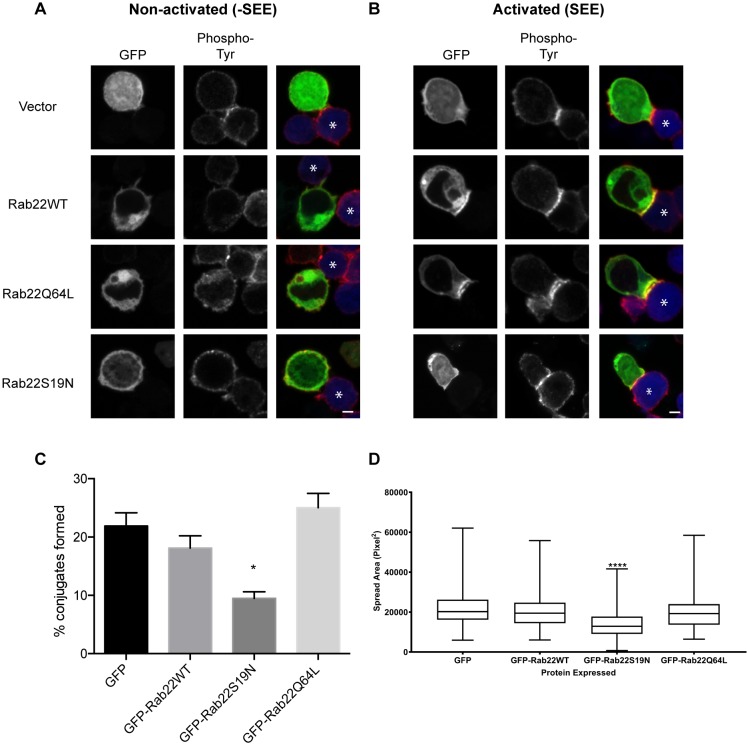
Rab22 polarizes to the immunological synapse and is required for the formation of stable T cell–APC conjugates. Jurkat cells were transfected with GFP-tagged Rab22WT, Rab22Q64L or Rab22S19N. Jurkat cells were then incubated with APCs or with APCs that had been exposed to SEE. Cells were fixed and phosphorylated tyrosine was detected with antibodies. (A) The non-activated control (-SEE) shows no polarization of Rab22 at the site of contact with the APC and phosphorylated tyrosine is low. (B) In the activated sample (+SEE), Rab22WT and Rab22Q64L polarize toward the site of contact and colocalize with phosphorylated tyrosine on the plasma membrane. Rab22S19N does not polarize toward the site of contact and reduces phosphorylated tyrosine at the site of contact. Asterisks mark APCs. Scale bars: 3 µm. (C) Cells expressing GFP or GFP-tagged Rab22 were incubated with APCs that had been exposed to SEE. After 1 h, cells were fixed and conjugate formation was quantified by using imaging flow cytometry. Shown are mean±s.d. from three experiments. *P<0.05 (unpaired t-test). (D) Jurkat cells expressing GFP-tagged Rab22WT, Rab22Q64L or Rab22S19N, or GFP alone, were plated on coverslips coated with antibodies against the T cell receptor (anti-CD3 antibody) for 3 min and then fixed and stained with a plasma membrane cell mask. The area the cell spread was quantified by using the image analysis software package Metamorph. Data is presented as box-and-whisker plots representing the median and the 25th and 75th percentiles. Whiskers show the minimum and maximum. n>150 cells over at least three independent experiments. ****P<0.0001 (one-way Anova with Kruskal–Wallis test).

