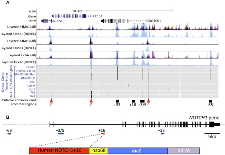
Fig. 1.
The human NOTCH1 locus contains multiple putative endothelial enhancers. (A) The human NOTCH1 locus from UCSC ENCODE Genome Browser (http://genome.ucsc.edu). Human umbilical vein endothelial cell (HUVEC)-specific H3me1 and H3K27ac (enhancer associated) and H3K4me3 (promoter associated) peaks are indicated in light blue [both in the separate HUVEC and combined tracks (denoted as ‘all’)]; other colours indicate H3K4me1, H3K4me3 and H3K4ac peaks specific to non-endothelial cell lines: GM12878 cells (red), H1-hESC cells (yellow), HSMM cells (green), K562 cells (purple) NHEK cells (lilac) and NHLF cells (pink). INPP5E and SEC16A are shown (blue text) in addition to NOTCH1 (black text). DNase I digital hypersensitive hotspots are indicated by black vertical lines on grey (HUVEC, HMVEC-dBl-Ad, HMVEC-dBl-Neo and HMVEC-LBl, which are all different endothelial cell types). The four NOTCH1 putative enhancer regions (black bars) were identified by high levels of HUVEC-specific H3K4me1 and H3K27ac associated with endothelial cell-specific DNaseI hypersensitivity hotspots. P, putative promoters identified by H3K4me3. (B) The human NOTCH1 gene (top, in 5′ to 3′ orientation with putative enhancers indicated) and the NOTCH1+16hsp transgene (bottom).

