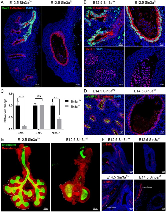
Fig. 4.
Sin3a is required for branching morphogenesis and cell fate in the developing lung. (A,B) Representative immunofluorescence staining of E12.5 embryonic lungs from Sin3af/f mutant and Sin3af/+ littermates showing Sox2 and E-cadherin (A), Sox9, E-cadherin (B, top) and Nkx2.1 (B, top). (C) Quantitative real-time PCR expression data for Sox2, Sox9 and Nkx2.1 for E12.5 embryonic lungs of Sin3af/f mutant and Sin3af/+ littermates. (D) Representative immunofluorescence staining of E14.5 embryonic lungs from Sin3af/f mutant and Sin3af/+ littermates showing proSP-C and E-cadherin. (E) Whole-mount confocal image of E12.5 embryonic lungs from Sin3af/f mutant and Sin3af/+ littermates. Dashed line delineates the mesoderm border of the right lobes. (F) Representative immunofluorescence staining of E12.5 and E14.5 embryonic lungs from Sin3af/f mutant and Sin3af/+ littermates showing spatial localization of α-SMA. Significance determined by Student's t-test; ns, not significant; **P<0.01, ****P<0.0001.

