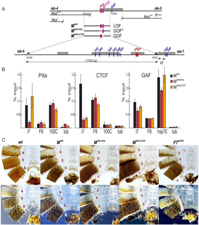
Fig. 4.
Role of Pita- and CTCF-binding sites in Mcp insulator function. (A) Schematic of the Mcp boundary and replacement fragments used for the insertion at F7attP50. The dCTCF-binding site is shown as magenta ovals. Bold black half-arrows (marked i7) below the map indicate primers that were used for ChIP experiments. Hypersensitive sites are shown as gray boxes. GAF- and Pita-binding sites are designated as blue and red ovals, respectively. (B) Binding of Pita, dCTCF and GAF at the PREiab-7 region in pupae of tested transgenic lines (M340, M340ΔPita and M340ΔCTCF). The results of ChIPs are presented as percentages of the input DNA. The 100C region was used as a positive control for Pita and hsp70 for GAF binding, the F8 region was used as a positive control for dCTCF and GAF binding, and the γTub37C-coding region was used as a negative control for binding. Error bars indicate standard deviations of triplicate PCR measurements from two independent biological samples of chromatin. (C) Morphology of the abdominal segments of the M340 mutant males (bright field, top row; dark field, bottom row). In M340 homozygous males, A6 is completely transformed into A5. The phenotypic effects are the same as in the case of Pita×5. M340ΔPita and M340ΔCTCF males demonstrate a mixed gain- and loss-of-function transformation of A6 segment. Phenotypes of M340 mutant females can be seen in Fig. S3.

