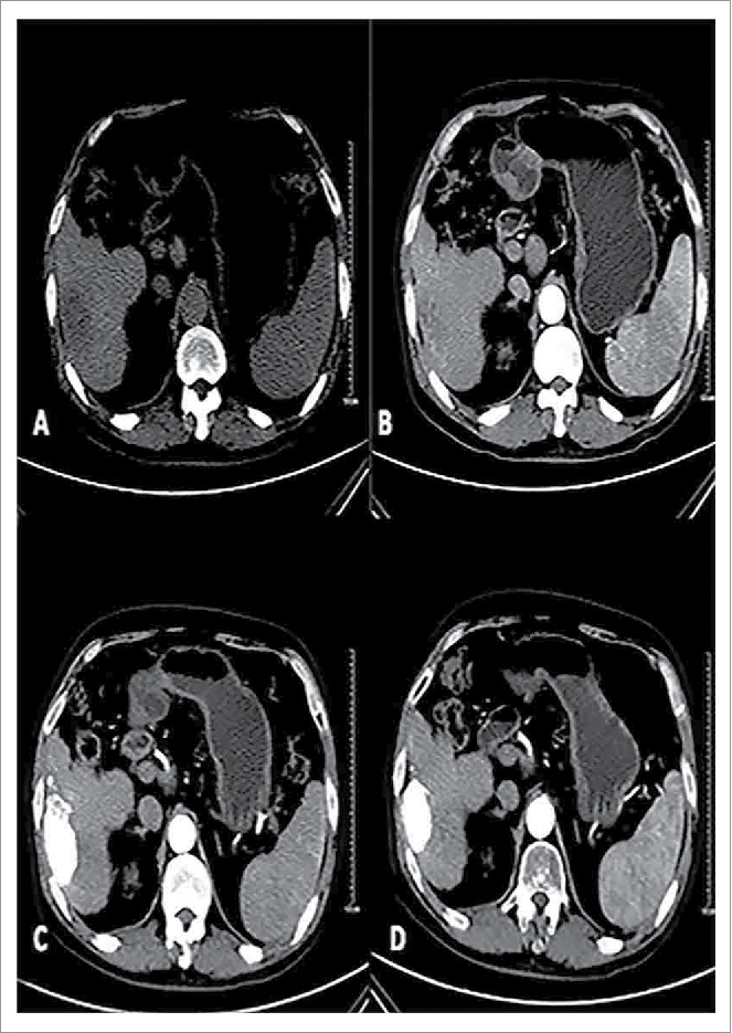
Figure 1.

Male patients, 57, primary liver cancer. Patients with preoperative CT scan of the right liver lobe low-density lesions, boundary is not clear, Size of 7.0 * 3.8 cm (A). Enhanced CT of the right liver lobe lesions early uneven arterial enhancement (B).With conventional therapy and oral path for 3 months after, the CT in tumor iodine oil deposits are good in the oven, enhanced scan did not see lesions.Curative effect evaluation for CR (C); Follow-up of 12 months after treatment tumors had the previous narrow (5.0× 2.7 cm), enhanced scan within tumors had no reinforcement (D).
