. 2017 Jun 29;6:e25093. doi: 10.7554/eLife.25093

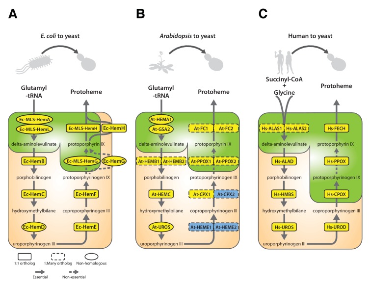
Figure 6. Yeast heme biosynthesis pathway enzymes can be successfully replaced by orthologs or analogs from bacteria, plants, and humans, in spite of alterations to subcellular localization.

Enzymatic steps of extant bacterial and eukaryotic heme biosynthesis pathways are identical save for the starting metabolites and conversion to delta-aminolevulinate; bacteria also exhibit non-orthologous gene displacement of several enzymes. Heme biosynthesis occurs in the bacterial cytoplasm and inner membrane, the human and yeast in mitochondria and cytoplasm, and the plant in chloroplast and cytoplasm. In spite of these localization changes over evolution, most of the defects in growth rate and viability conferred by heme pathway mutations in yeast can be complemented by introduction of the corresponding (A) bacterial genes, (B) plant genes (except for At-HemE), and (C) human genes. Yellow indicates a replaceable gene, blue non-replaceable.

DOI: http://dx.doi.org/10.7554/eLife.25093.017

Figure 6.

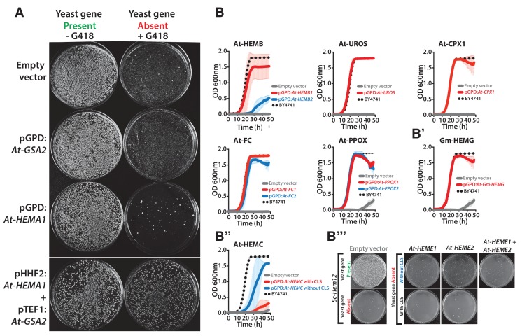
Figure 6—figure supplement 1. Heme biosynthesis genes from Arabidopsis thaliana and Glycine max generally efficiently replace their counterparts in yeast, except in the case of ΔSc-Hem12.

Figure 6—figure supplement 1.

(A) Expression of heme pathway genes from Arabidopsis thaliana, At-HEMA1 or At-GSA2, individually cannot complement the lethal growth defect of the deletion of Sc-hem1 gene in yeast. Co-expression of At-HEMA1 and At-GSA2 rescued the growth defect of Sc-hem1 gene deletion in yeast. (B) Haploid yeast gene deletion strains carrying plasmids expressing functionally replacing Arabidopsis (red or blue solid-lines) and (B’) Glycine max (Gm-HEMG) heme pathway genes (red solid-line) generally exhibit comparable growth rates to the wild type parental yeast strain BY4741 (black dotted-line) as grown in magic marker liquid medium in the presence of G418 (200 μg/ml). (B’’) Native At-HEMC with chloroplast localization signal (CLS) showed poor replaceability in yeast (red solid-line). Removal of the CLS from At-HEMC allowed efficient rescue of the corresponding yeast gene deletion, ΔSc-Hem3 (blue solid-line). (B’’’) However, neither the expression of Arabidopsis proteins At-HEME1 or At-HEME2 (with or without CLS) alone nor their co-expression could functionally rescue the corresponding yeast gene deletion, ΔSc-Hem12. Wild type BY4741 haploid strain is plotted for comparison (black dotted-line). Strains carrying empty vector were used as controls (grey solid-line). Mean and standard deviation plotted with N = 3.
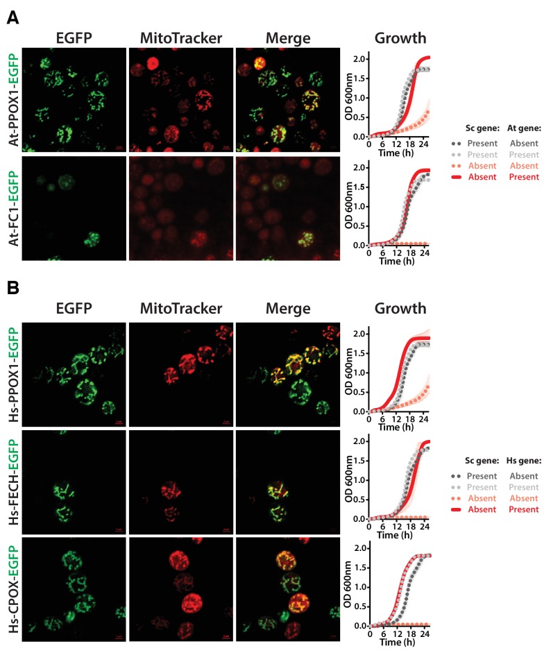
Figure 6—figure supplement 2. Heme biosynthesis enzymes normally localized to plant chloroplasts or human mitochondria localize to the mitochondria when expressed in yeast.

Figure 6—figure supplement 2.

(A) EGFP-tagged penultimate At-PPOX1-EGFP and ultimate At-FC1-EGFP proteins localize to mitochondria in yeast. Green fluorescence proteins co-localized with Mitotracker red-stained mitochondria. In certain cases, At-FC1-EGFP formed aggregates. Expression of EGFP-tagged plant genes, At-PPOX1-EGFP and At-FC1-EGFP (red solid-line), efficiently rescue the growth defect of the corresponding yeast gene deletions (pink dotted-line). The over-expression of the tagged proteins is not toxic to the wild type yeast strain (grey dotted-line). The growth rescue by plant genes is as efficient as the wild type BY4741 yeast strain (black dotted-line). Mean and standard deviation plotted with N = 3. (B) The EGFP-tagged last three heme pathway genes from humans localize to mitochondria in yeast. The green fluorescence co-localized with the Mitotracker red-stained mitochondria in yeast. Expression of EGFP-tagged human genes, Hs-PPOX-EGFP, Hs-FECH-EGFP and Hs-CPOX-EGFP (red solid-line), efficiently rescue the growth defect of the corresponding yeast gene deletions (pink dotted-line). The over-expression of the tagged proteins is not toxic to the wild type yeast strain (grey dotted-line). The growth rescue by the human genes is as efficient as the wild type BY4741 yeast strain (black dotted-line). Mean and standard deviation plotted with N = 3.
Figure 6—figure supplement 3. Human heme biosynthesis genes efficiently replace their yeast counterparts.

Figure 6—figure supplement 3.

Functional replacement of human genes in yeast. (A) Expression of Hs-UROS in Sc-hem4 heterozygous diploid deletion yeast strain resulted in toxicity post-sporulation as seen by the lack of growth on either magic marker agar medium with (yeast gene present) or without G418 (yeast gene absent). (B) This toxicity was relieved by replacing the human Hs-UROS at the native yeast locus. Growth curve of the humanized yeast Sc-hem4Δ::Hs-UROS strain (red-solid line) showed comparable growth to the wild type yeast BY4741 (black dotted-line). (C) Expression of human Hs-UROD (a human orfeome clone with G303V mutation) in Sc-hem12 heterozygous diploid deletion yeast strain did not complement the growth defect of the yeast gene as shown by plating the post sporulation mix on magic marker medium with or without G418. Reverting the sequence to the wild type Hs-UROD gene resulted in efficient rescue of the growth defect of the corresponding yeast gene. (D) Expression of human genes, Hs-PPOX, Hs-UROD, Hs-ALAS1 (red solid-line) and Hs-ALAS2 (blue solid-line), efficiently rescue the growth defect of the corresponding yeast gene deletions (grey solid-line), Sc-hem14 and Sc-hem1, respectively. The rescue was largely comparable to the wild type BY4741 yeast strain (black dotted-line). Strains carrying empty vector were used as controls (grey solid-line). Mean and standard deviation plotted with N = 3.