
Figure 1.
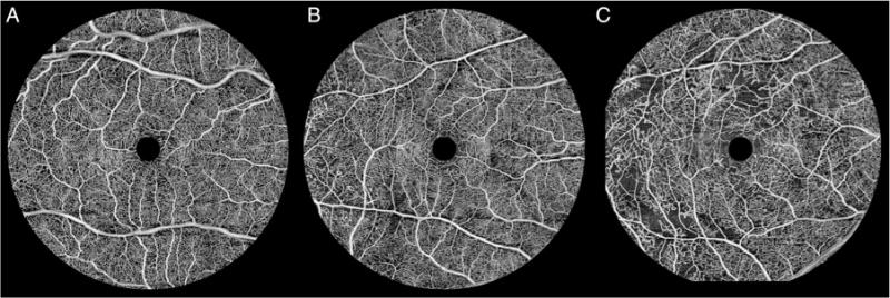
En face ocular coherence tomography-based microangiography images of macular capillary networks for the following diabetic retinopathy groups: none–mild non-proliferative diabetic retinopathy (A), moderate–severe non-proliferative diabetic retinopathy (B) and proliferative diabetic retinopathy (C).