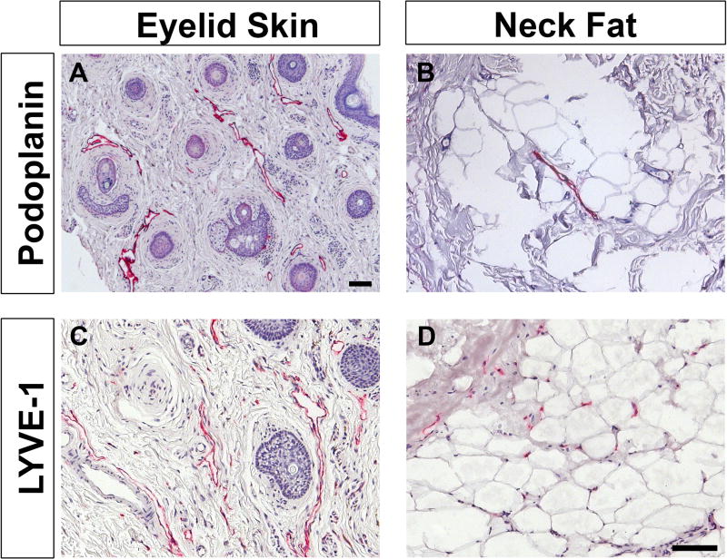
Figure 1. Immunohistological characterization of control specimens, eyelid skin and subcutaneous fat obtained from the neck.
Localization of podoplanin and LYVE-1 (red) confirm the presence of lymphatic vessels in these samples, as expected, and proves the utility of these markers. Samples are counterstained with hematoxylin (blue). Scale bar = 100 µm

