Figure 5.

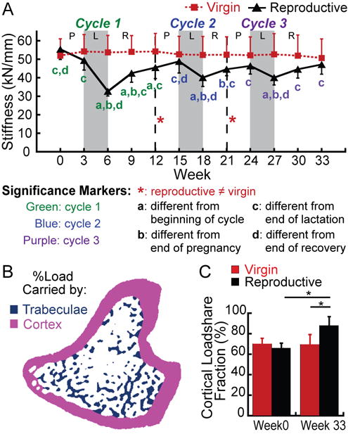
(A) Changes in whole-bone stiffness at the proximal tibia over 3 cycles of pregnancy (P), lactation (L), and post-weaning recovery (R) in reproductive rats (black triangles), and virgins (red squares). Letters indicate significant differences (p<0.05) among time points for each cycle in the reproductive group (no significant changes over time were found for virgin rats). Red # indicates a trend towards a difference between reproductive and virgin rats at the end of the first reproductive cycle (p<0.1). (B) Schematic illustrating the separation of cortical (pink) and trabecular (blue) compartments in order to determine the percentage of the load carried by the trabecular and cortical bone (load-share fraction). (C) Percentage of load carried by the cortical bone at baseline and after 3 reproductive cycles in virgin and reproductive rats. *: p<0.05.
