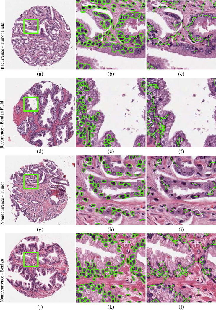
Fig. 1.
Prostate tissue microarray cores (a, d, g, j) corresponding to patients who experienced (a–f) recurrence and (g–l) no recurrence. Automated segmentation defines nuclear boundaries and locations from tumor and benign field tissue cores, characterizing (b, e, h, k) nuclear shape and (c, f, i, l) nuclear subgraphs describing local nuclear architecture.

