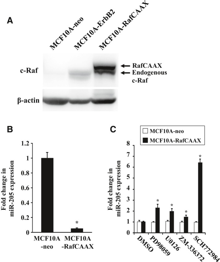
Figure 2.

Expression of miR‐205 in MCF10A‐RafCAAX cells treated with ERK inhibitor. (A) Western blot analysis of MCF10A‐RafCAAX, MCF10A‐ErbB2, and MCF10A‐neo cells with anti‐Raf‐1 antibodies. β‐Actin was used as a control for loading. (B) Real‐time quantitative RT‐PCR analysis of miR‐205 expression in MCF10A‐RafCAAX and MCF10A‐neo cells. Data are mean ± SEM of three independent experiments. (C) Real‐time quantitative RT‐PCR analysis of miR‐205 expression in MCF10A‐neo and MCF10A‐RafCAAX cells treated with MEK inhibitor U0126 (10 μm) or PD98059 (20 μm), Raf1 kinase inhibitor ZM‐336372 (1 μm), or ERK inhibitor SCH772984 (1 μm) for 48 h. Data are mean ± SEM of three independent experiments. *P < 0.01 by Student's t‐test compared with MCF10A‐neo.
