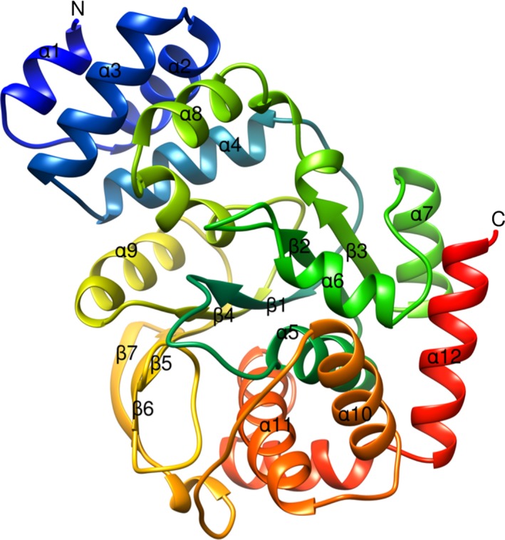
Figure 7.

Ribbon diagram of X‐ray crystal structure of TkTrpD. Only one subunit of the homodimer is shown, with the amino acid chain colored from blue at the N terminus to red at the C terminus. Each subunit consists of a small α‐helical domain containing four helices (α1, α2, α3, and α4) and a larger C‐terminal α/β domain with a central β‐sheet containing seven β‐strands (six parallel and one antiparallel) surrounded by eight α‐helices. Figure was created using UCSF Chimera.
