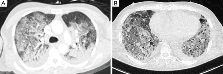
Figure 2.
Computed tomography of two patients with ARDS. (A) Patient with a severe ARDS in early phase, showing diffuse bilateral infiltrates and small dorsal consolidated areas; (B) patient with a fibrotic pattern developed in the late phase of an ARDS, showing diffuse infiltrates with areas of ‘honeycomb’ pattern and left pleural effusion. The scans were obtained on the same patients and same day as in Figure 1. ARDS, acute respiratory distress syndrome.

