| A |
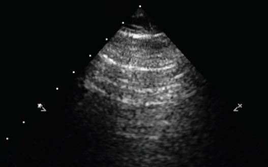 |
Reverberation artefacts parallel to the pleural line (A-lines) |
Expression of a physiologically aerated lung when accompanied by the sliding sign of the pleural layers, but could also be present in hyper-inflated regions—normal lung aeration |
| B1 |
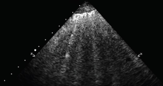 |
Multiple vertical artefacts (B-lines) |
Correlated to extra vascular lung water content, absent in normal lung, hallmark of the alveolo-interstitial syndrome—moderate loss of aeration |
| B2 |
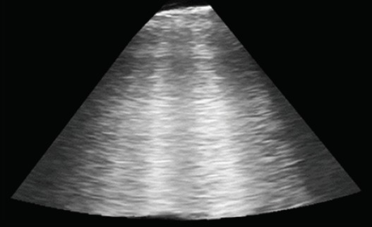 |
Multiple, coalescent B-lines |
Further progression of the B1 pattern—severe loss of aeration |
| C |
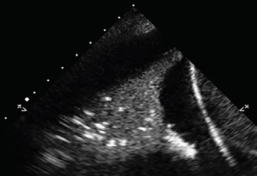 |
Real image, not artefact with tissue-like echogenicity, or shred sign (typical of alveolar consolidation) |
Visible when consolidations are extended to the pleura—collapsed or consolidated lung tissue |
