Figure 5.

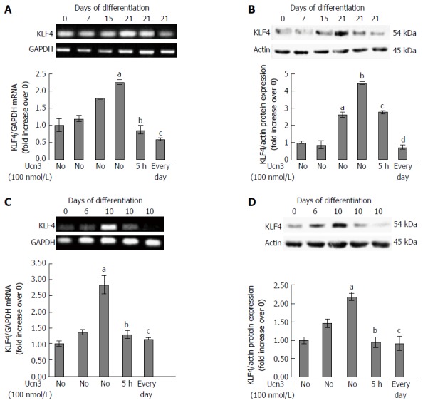
Down-regulation of KLF4 mRNA and protein expression following corticotropin releasing factor receptor 2 signaling. A: Detection of KLF4 mRNA expression by RT-PCR during the kinetic of Caco-2 cell differentiation and after acute (5 h) or chronic (every day) exposure to 100 nmol/L Ucn3 of 21 d differentiated cells. GAPDH served as a housekeeping control. Quantification of KLF4 mRNA from RT-PCR assays (lower panel). Data were expressed as fold increase of KLF4/GAPDH mRNA levels of differentiated (D7, D15, D21) vs undifferentiated cells (D0). Data represents means of three different experiments ± SEM. a,bP < 0.001 vs undifferentiated Caco-2 cells (D0); cP < 0.001 vs differentiated Caco-2 cells (D21). B: Detection of KLF4 protein expression by western blot during the kinetic of Caco-2 cell differentiation and after acute (5 h) or chronic (every day) exposure to 100 nmol/L Ucn3 of 21 d differentiated cells. Actin served as a loading control. Lower panel: Quantification of KLF4 protein levels from western blot analyses. Data were expressed as fold increase of KLF4/actin protein levels of differentiated (D7, D15, D21) vs undifferentiated cells (D0). Data represents means of three different experiments ± SEM. a,bP < 0.001 vs undifferentiated Caco-2 cells (D0); c,dP < 0.001 vs differentiated Caco-2 cells (D21). C: Detection of KLF4 mRNA expression by RT-PCR during the kinetic of HT-29 cell differentiation and after acute (5 h) or chronic (every day) exposure to 100 nmol/L Ucn3 of 10 d differentiated cells. GAPDH served as a housekeeping control. Quantification of KLF4 mRNA from RT-PCR assays (lower panel). Data were expressed as fold increase of KLF4/GAPDH mRNA levels of differentiated (D6 and D10) vs undifferentiated cells (D0). Data represents means of three different experiments ± SEM. Data represents means of three different experiments ± SEM. aP < 0.001 vs undifferentiated HT-29 cells (D0); bP < 0.05 vs early differentiated HT-29 cells (D10), cP < 0.01 vs D10. D: Detection of KLF4 protein expression by western blot during the kinetic of HT-29 cell differentiation and after acute (5 h) or chronic (every day) exposure to 100 nmol/L Ucn3 of 10 d differentiated cells. Actin served as a loading control. Lower panel: Quantification of KLF4 protein levels from western blot analyses. Data were expressed as fold increase of KLF4/actin protein levels of differentiated (D6 and D10) vs undifferentiated cells (D0). Data represents means of three different experiments ± SEM. aP < 0.001 vs undifferentiated HT-29 cells (D0); b,cP < 0.001 vs early differentiated HT-29 cells (D10).
