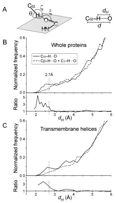
Figure 1.
(A) Definition of the geometrical parameters of the Cα—H⋅⋅⋅O hydrogen bond. Nomenclature according to Derewenda et al. (14). The ideal values (14, 18) are as follows: H—O distance, dH ≤ 2.7 Å; Cα—O distance, d ≤ 3.8 Å; Cα—H—O angle, ζ = 180°; H—O—C angle, ξ = 120°; elevation angle, θ = 0° (angle between the Cα—H vector and the amide plane). (B) Distribution of hydrogen to acceptor distances (dH) for all Cα—H donor groups (solid line), compared with that of Cβ—H + Cγ—H groups (dashed line), in the 11 membrane proteins structures. The ratio between the two curves is also shown (Cα/Cβγ). The control set is similarly sized and is composed by the Cβ—H groups of all Ile, Leu, Val, Met, Phe, and Tyr residues and the Cγ—H of Leu and Cγ1—H of Ile. Contacts below 2.7 Å have an overall frequency of occurrence that is 3 times higher for Cα donors than for the Cβγ control set. (C) Analogous distribution as in B, but limited to the subset of residues in helical transmembrane segments.

