
Fig. 3.
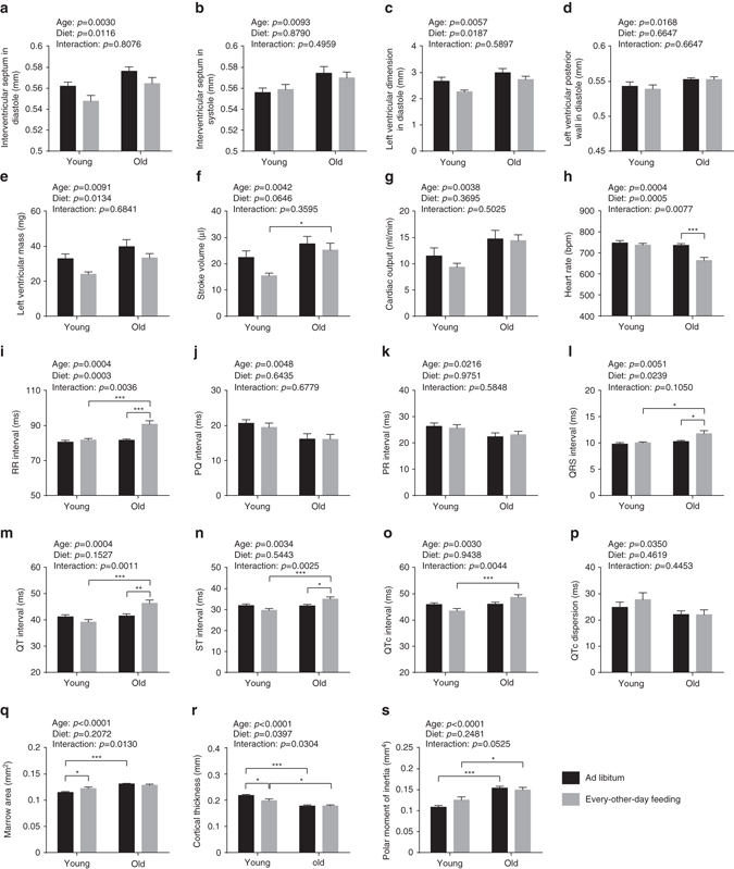
Cardiological analyses and assessment of bone structure. a–g Ventricular dimensions and functions were examined with transthoracic echocardiography (two-way ANOVA; young + AL: n = 10, young + EOD: n = 9, old + AL: n = 11, old + EOD: n = 11). Dimensions of a interventricular septum in diastole, b interventricular septum in systole, c left ventricle in diastole, and d left ventricular posterior wall in diastole were determined. e Mass of left ventricle, f stroke volume, and g cardiac output were calculated. h–p ECGs were recorded from conscious mice (two-way ANOVA; young + AL: n = 10, young + EOD: n = 9, old + AL: n = 11, old + EOD: n = 11). h HR, i RR interval, j PQ interval, k PR interval, l QRS interval, m QT interval, n ST interval, o QTc interval, p QTc dispersion. q–s Bone architecture was examined using micro-CT of distal tibia (two-way ANOVA; young + AL: n = 16, young + EOD: n = 9, old + AL: n = 17, old + EOD: n = 9). q Marrow area, r cortical thickness, s polar moment of inertia. AL ad libitum, EOD every-other-day feeding. Data are shown as mean ± SEM. Full data sets of echocardiography, electrocardiography, and micro-CT assessments are presented in Supplementary Tables 10–12