
Fig. 5.
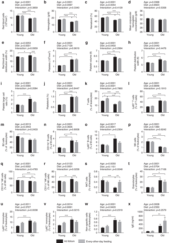
Hematological and immunological analyses. a–j Number and size of the different blood cell types were determined using a hematology analyzer (two-way ANOVA; young + AL: n = 16, young + EOD: n = 16, old + AL: n = 20, old + EOD: n = 22). a RBC count, b hemoglobin concentration, c HCT, d MCHC, e RDW, f platelet count, g platelet volume, h PDW, i large cell ratio of platelets (>12 fl), and j PCT. k–w Peripheral blood leukocytes were analyzed using a ten-color flow cytometer (two-way ANOVA; young + AL: n = 16, young + EOD: n = 16, old + AL: n = 20, old + EOD: n = 22). Only CD45-positive cells were included in the analysis. k T cells (CD3+CD5+), l Ly6C+ T cells, m B2 cells (CD5−CD19+B220+), n CD11b+ B cells (CD19+B220+), o Ly6C+ B cells (CD19+B220+), p natural killer (NK) cells (CD3−CD5−NKp46+ and/or NK1.1+), q CD11b+ NK cells, r CD11c+ NK cells, s NKT cells (CD3+CD5+NKp46+ and/or NK1.1+), t granulocytes (CD11b+Ly6G+), u Ly6C+ monocytes (CD11b+), v Ly6C− monocytes (CD11b+), w non-specific cells, x plasma concentration of IgE immunoglobulins. AL ad libitum, EOD every-other-day feeding. Data are shown as mean ± SEM. Full data sets of hematological and immunological analyses are presented in Supplementary Tables 14 and 15