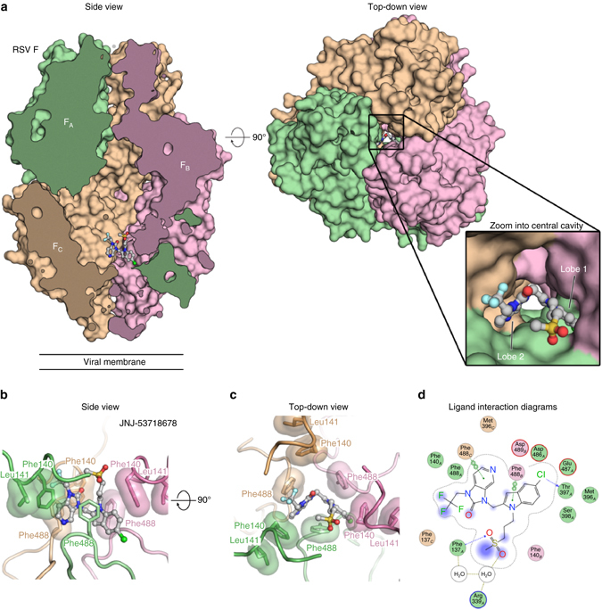
Fig. 1.

JNJ-53718678 binds to a threefold-symmetric cavity in prefusion RSV F. a Side and top-down views of the crystal structure of JNJ-53718678 bound to prefusion RSV F. RSV F is shown as a molecular surface with the three identical protomers each shown in a different color (FA, green; FB, pink; and FC, tan). JNJ-53718678 is shown as ball-and-stick representation with carbon atoms colored in grey, nitrogen atoms in blue, oxygen atoms in red, chlorine atom in dark green, fluorine atoms in light blue, and sulfur atoms in orange. For clarity, part of the front hemisphere in the side view panel was removed. The inset provides a zoomed view of the binding of JNJ-53718678 into the central cavity. b, c Side b and top c views for JNJ-53718678 bound to prefusion RSV F. Each RSV F protomer is again shown in a different color corresponding to the colors in a, and hydrophobic side chains are shown with transparent molecular surfaces. JNJ-53718678 is shown as ball-and-stick representation with colors of atoms corresponding to the colors in a. d 2D ligand-interaction diagram generated in Molecular Operating Environment. Coloring of circles refers to the green (FA), pink (FB), and tan (FC) protomers. Red and blue circled outlines represent negatively and positively charged groups, respectively. Bonds with RSV F main chain and side chain atoms are shown as blue and green dashed lines, respectively, and water-mediated interactions are shown as light brown dashed lines. When present, arrowheads point toward the acceptor
