Figure 4.

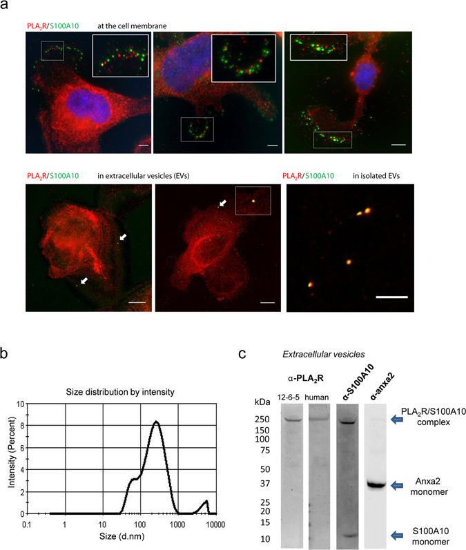
PLA2R is found in vesicles secreted by podocytes. (a) Co-localisation of PLA2R/S100A10 at the cell surface and in vesicles. Podocytes cultured on coverslips were fixed and co-stained using antibodies against S100A10 (green) and PLA2R (red) mAb 12-6-5. Merged images demonstrate some regions of overlap (yellow) between the PLA2R and S100A10 at the cell membrane (top panel) and in vesicles (left bottom panels). Scale bars, 10 μm. Purified extracellular vesicles (EVs) secreted from podocytes were incubated on poly-D-lysine coated glass slide and co-stained for PLA2R and S100A10 showing positive staining for both proteins. (b) Representative size distribution by intensity profile of extracellular vesicles (EVs) measured by Dynamic Light Scattering (DLS). EVs intensity distributions reveal two characteristic peaks occurring at ~60 nm and ~200 nm. These two values are in agreement with the average values reported in literature for exosomes and microvesicles, respectively. (c) Vesicles characterisation. Isolated EVs from serum-free conditioned cell culture media of differentiated podocytes were analysed by western blotting using anti-PLA2R, anti-S100A10 and anti-Anxa2 antibodies. A ~250 kDa band was detected by both anti-PLA2R and anti-S100A10 antibodies corresponding to the tightly associated PLA2R/S100A10 complex. Anxa2 was also detected in the EVs fraction as a monomer around ~37 kDa suggesting it has dissociated from the complex under denatured conditions.
