
Figure 2.
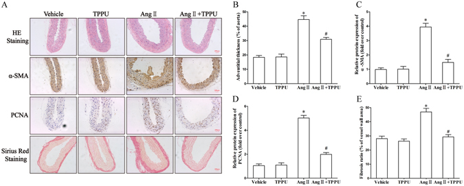
TPPU administration attenuated Ang II-induced adventitial remodeling in C57BL/6 J mice. (A) Vessel sections were performed with hematoxylin and eosin staining, sirius red staining, and immunohistochemical staining of α-SMA and PCNA. (B) Quantitative analysis of adventitial thickness. (C) Quantitative analysis of adventitial α-SMA expression. (D) Quantitative analysis of adventitial PCNA expression. (E) Quantitative analysis of adventitial fibrosis. Data were expressed as Mean ± SEM, n = 8 mice for each group. *p < 0.05 vs. Vehicle, #p < 0.05 vs. Ang II.