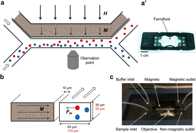
Figure 1.

Design of microfluidic sorting device. The microfluidic device is shown from above (a,a’) and as a cross section trough the channels (b). A ferrofluid is filled in the side channel distanced 10 µm from the sorting channel (a’). In the presence of an external magnetic field (thick arrows, H) the nanoparticles in the ferrofluid align to magnetize the ferrofluid (thin arrows, M) and create a gradient field. This results in an attractive force (F m) acting on magnetic cells (red), whereas non-magnetic cells (blue) continue in the sample flow (a and b). The small magnetotactic bacteria were tested in a sorting channel with reduced dimensions (black) compared to dimensions used for macrophages (red). The microfluidic chip (a’) is placed inside a chip holder to ensure stable connection of the tubing and placed under a fluorescent microscope for online monitoring of the separation process (c).
