Fig. 3.

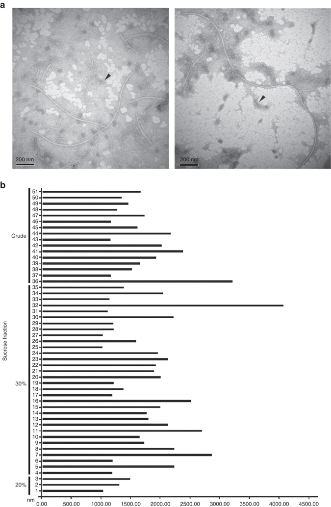
Representative transmission electron microscopy (TEM) images of virus-like particles extracted from Colletotrichum camelliae strain LT-3-1 and a histogram of the sizes of particles longer than 1000.0 nm. a Representative virus-like particles extracted from strain LT-3-1 corresponding to the 30% fraction following sucrose gradient centrifugation. The arrows indicate empty particles that might have lost dsRNA. Scale bars, 200 mm. b A histogram of the sizes of particles longer than 1000.0 nm from strain LT-3-1 in crude extracts and in fractions corresponding to 30% and 20% sucrose fractions, respectively, following sucrose gradient centrifugation. The numbers on the vertical axis indicate numbers randomly assigned to virus-like particles
