Fig. 5.

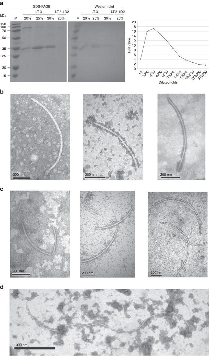
Western blot analysis and titer quantification of a polyclonal antibody against CcFV-1 P4 and TEM and immunosorbent electron microscopy (ISEM) analysis of virus particles. a SDS-PAGE (left panel) analysis of the proteins from strain LT-3-1 in 20–30% sucrose fractions after sucrose gradient centrifugation and from strain LT-3-1D2 in the 25% sucrose fraction; Western blot analysis of these proteins by SDS-PAGE (right panel) using the antibody against CcFV-1 P4 (PAb-P4). Titer quantification of PAb-P4 by indirect ELISA against the P4 protein from fractions after sucrose gradient centrifugation using PAb-P4 diluted from 50- to 512,000-fold. P/N, ratio of the absorbance values of the positive sample (CcFV-1-infected LT-3-1) against the negative sample (virus-free LT-3-1D2) at a wavelength of 450 nm. b, c Direct TEM and ISEM analysis of virus-like particles derived from 20% and 30% fractions following sucrose gradient centrifugation of strains LT-3-1 (b) and LT-3-1T2 (c), respectively. The ISEM images indicate that the virus-like particles are decorated by PAb-P4 at a 2000-fold dilution. Scale bars, 200 mm. d The longest virus-like particles in the crude extract decorated by PAb-P4 at a 8000-fold dilution, as observed by ISEM. Scale bar, 1000 mm
