Figure 1.

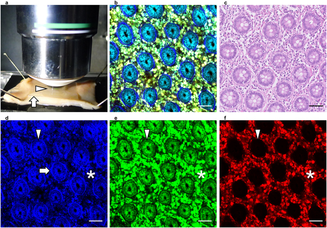
Non-labeling MPM imaging of human normal colorectal mucosa. (a) Overview of the imaging approach. Colorectal tissue was placed on a rubber plate with the mucosal surface facing upwards using small pins and was overlaid with a coverslip (arrow) to form a drop of water (arrow head). (b) MPM imaging of human normal colorectal mucosa. (c) HE staining of normal colorectal mucosa, using the same sample as in (b). (d–f) Images obtained using a 417/60 nm filter (d), 480/40 nm filter (e), and 629/56 nm filter (f) of the samples used in (b). Fibrous structures (arrow), epithelial cells (arrow head), and immune cells (asterisk) showed different color patterns. Bar: 50 µm.
