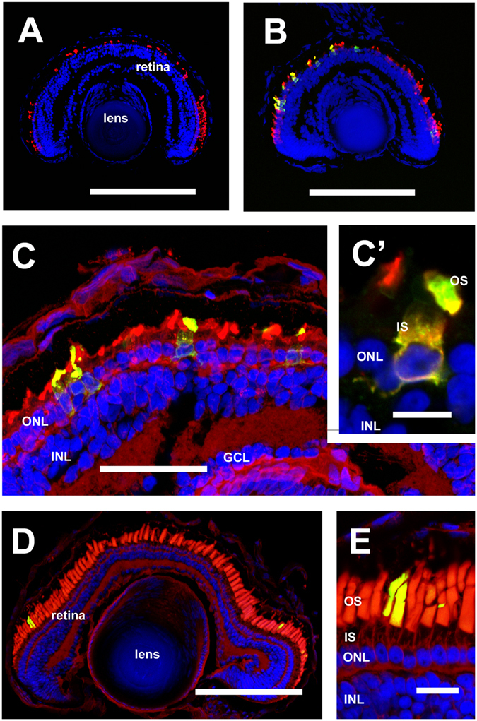
Figure 7.

HDR mediated gene alterations assessed by confocal microscopy: (A) RD in an animal edited using sgRNA rhosg4 (B,C,C’) images derived from an animal in which rhosg4 was used to direct targeted insertion of eGFP into rho.L. A number of eGFP-positive rods are present, while other cells of the retina and lens do not show eGFP expression. Significant RD is present (D,E) images derived from an animal with targeted mutation of residue M13→F in rho.L, and stained with anti-mammalian rhodopsin (2B2). There is no identifiable RD. (A–C) Green: eGFP. (D,E) green: anti-mammalian rhodopsin (2B2). (A,B) Red: anti-rod transducin. C–E: Red: wheat germ agglutinin. (A–E) Blue: Hoechst 33342. OS: outer segments. IS: inner segments. ONL: outer nuclear layer. INL: inner nuclear layer. GCL: ganglion cell layer. (A,B,D) Bar = 200 µm. (C) Bar = 50 µm. (C’) Bar = 10 µm. (E) Bar = 20 µm. Panels C and C’ are confocal projections derived from 10 confocal sections.
