Figure 2.

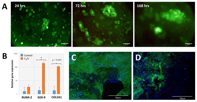
(A) Human adipose derived pericyte cultured within Fmoc-F2/S hydrogels. Cells were encapsulated in F2/S hydrogels and maintained in unsupplemented basal media for up to 1 week. Cells were checked for viability by fluorescence detection of Syto 10 (green) for live cells and ethidium homodimer-1 (red) for dead cells after 1, 3 and 7 days. (B) QRT-PCR analysis for gene expression of pericyte cells cultured within 15.5 kPa Fmoc-F2/S hydrogels. Cells were assessed for the production of chondrogenic biomarkers RUNX-2, SOX-9 & type II collagen (COL2A1) after one week in culture. (C & D) Confocal microscopy images of immunofluorescently stained F2/S hydrogels cultured with pericytes for 28 days. Pericytes were checked for chondrogenic development by staining for aggrecan production (C) and type II collagen (D) both ascertained through green fluorescence. Cell populations are indicated by staining the cell nucleus with DAPI (blue). The images are mosaics of a 3 × 3 tile scan, each acquired from random positions of the hydrogel. Scale bar in A is 100 µm, in C & D is 50 µm. In B, Error bars denote the standard error where p < 0.05 as calculated using unpaired student t-test.
