Figure 2.

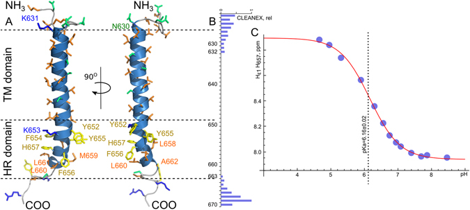
Spatial structure of TLR4-TMICL. (A) NMR-derived spatial structure of TLR4-TMICL in DMPG/DHPC q = 0.4 bicelles, pH 6.0, LPR 200. Residues in the hydrophobic part of the ICL region of the protein are indicated and painted according to their physical properties: Hydrophobic by orange, aromatic by yellow, polar by green and positively charged – by blue. (B) relative rates of the amide protons exchange with the solvent, as measured in the CLEANEX experiment26 for the TLR4-TMICL in DMPG/DHPC q = 0.4 bicelles at pH 7.2. (C) Dependence of the Hε1 chemical shift of H657 on the ambient pH which was used to determine the corresponding pKa. The fit of the obtained data to the equation (1) is shown by the red solid line.
