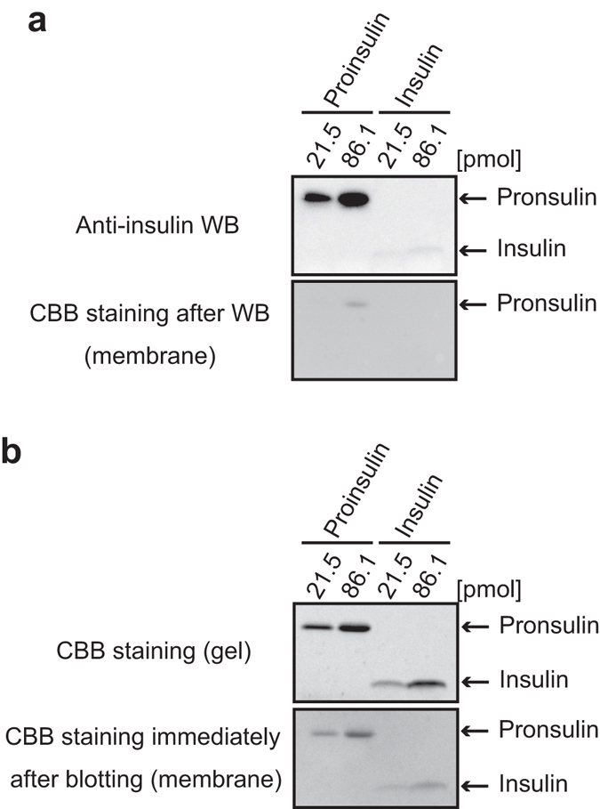
Figure 2.

During the general WB method, insulin is easily washed out from the blotted membrane compared with proinsulin. The indicated doses of proinsulin and insulin were separated by Tris/Tricine/urea SDS-PAGE. (a) WB was performed by using anti-insulin clone c27c9, which recognizes both proinsulin and insulin (upper panel). After WB detection, the membrane was stained with CBB R-250 (lower panel). In the condition, insulin was below detection limit of CBB staining of the membrane. (b) The PVDF membrane immediately after electro-blotting for WB (upper panel) was stained with CBB R-250. As another experiment, the separated gel was directly stained with CBB R-250 (lower panel).
