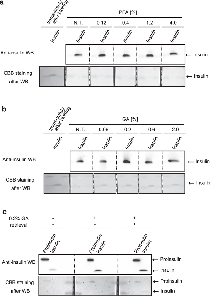
Figure 3.

Glutaraldehyde efficiently fixes insulin on the blotted membrane. (a,b) Reference standard of insulin (1 µg) was subjected to Tris/Tricine/urea SDS-PAGE. After electro-blotting, the one blotted membrane was separated into 1 slip for the control experiment (Coomassie brilliant blue [CBB] staining immediately after electro-blotting, labeled as “Immediately after blotting”) and 5 slips for the aldehyde treatment. Paraformaldehyde (PFA) (a) or glutaraldehyde (GA) (b) concentrations were verified as shown. N.T. indicates a control treatment. (c) Reference standards of proinsulin (200 ng) and insulin (125 ng) were subjected to Tris/Tricine/urea SDS-PAGE. After electro-blotting, the blotted membrane was separated into 3 slips to verify the effect of the retrieval step on insulin Western blotting (WB). Upper panels are the results of WB of insulin, and the lower panels are the results of CBB staining of the blotted membrane. To compare the effects of various conditions, WB images in each sub-part of the figure were captured at one time.
