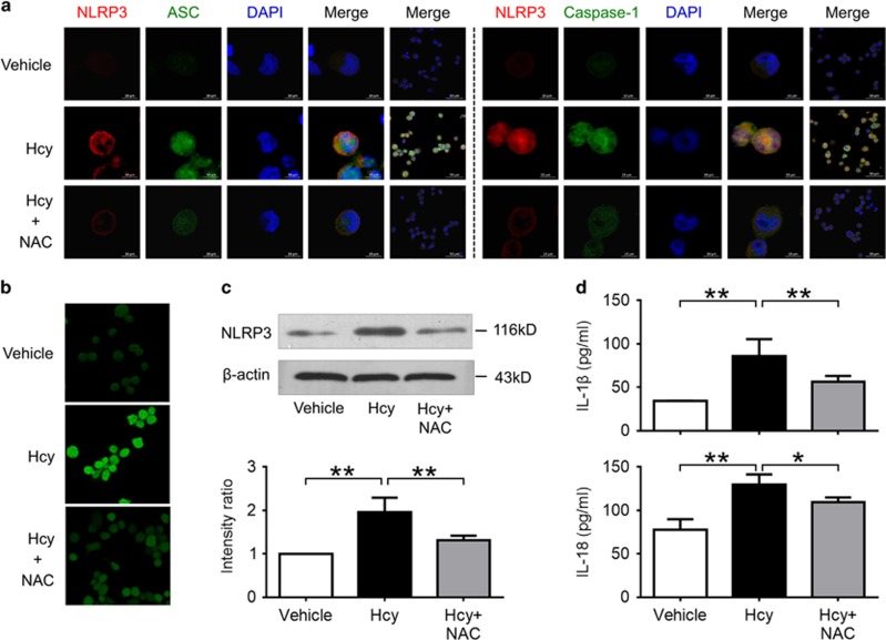
Figure 7.
Hcy activated NLRP3 inflammasomes in a ROS-dependent pathway. THP-1-differentiated macrophages were preincubated with NAC (50 mmol/l) for 1 h, then treated with 200 μmol/l Hcy for 0.5 h (b) or 24 h (a, c, d). Intracellular ROS levels were measured using a cell-permeable fluorescent probe, 2’7’-dichlorofluorescein diacetate (DCFH-DA). Data are representative of three independent experiments. (a) Representative confocal microscopic images showing colocalization of NLRP3 (red) with ASC (green), or NLRP3 (red) with caspase-1 (green) in macrophages. (b) Representative confocal microscopic images showing ROS accumulation in macrophages. (c) Representative blots and quantitative analysis of NLRP3 to β-actin. (d) Levels of IL-1β and IL-18 in cultured supernatant were measured by ELISA. Hcy, homocysteine; NAC, N-acetyl-l-cysteine. *P<0.05, **P<0.01.

