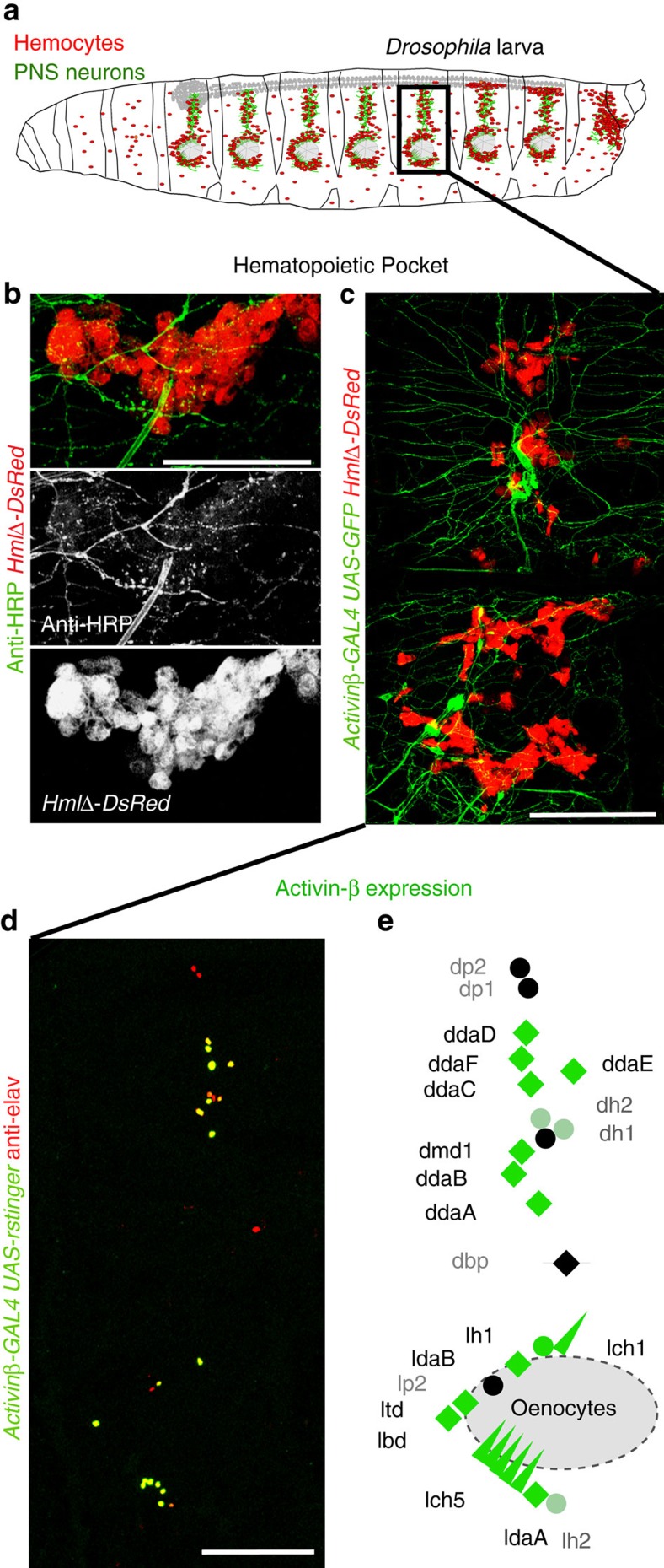
Figure 1. PNS sensory neurons produce Activin-β and are surrounded by hemocytes.
(a) Model of a Drosophila larva; hemocytes in red, neurons in green. Boxed area marks a HP. (b) Close-up of a lower part of a HP illustrating intricate neuronal extensions in areas of hemocytes; neurons (Anti-HRP, green), hemocytes (HmlΔ-DsRed, red), lateral patch area. Middle and lower panel show single channels. (c) Actβ expressing larval PNS neurons marked by reporter Actβ-GAL4; UAS-mCD8GFP (green), Actβ positive neurons colocalize with hemocytes marked by HmlΔ-DsRed (red). Fillet prep containing all tissue layers. (d) Actβ expression pattern in larval PNS neurons, Actβ-GAL4/+; UAS-r-stinger/+ (green), pan-neuronal anti-elav (red). Fillet prep containing all tissue layers. (e) Model of Actβ expression (green) in most multidendritic neurons (diamonds), chordotonal organs (triangles), and some external sensory neurons (circles). Scale bars, b, 50 μm; c,d, 100 μm.

