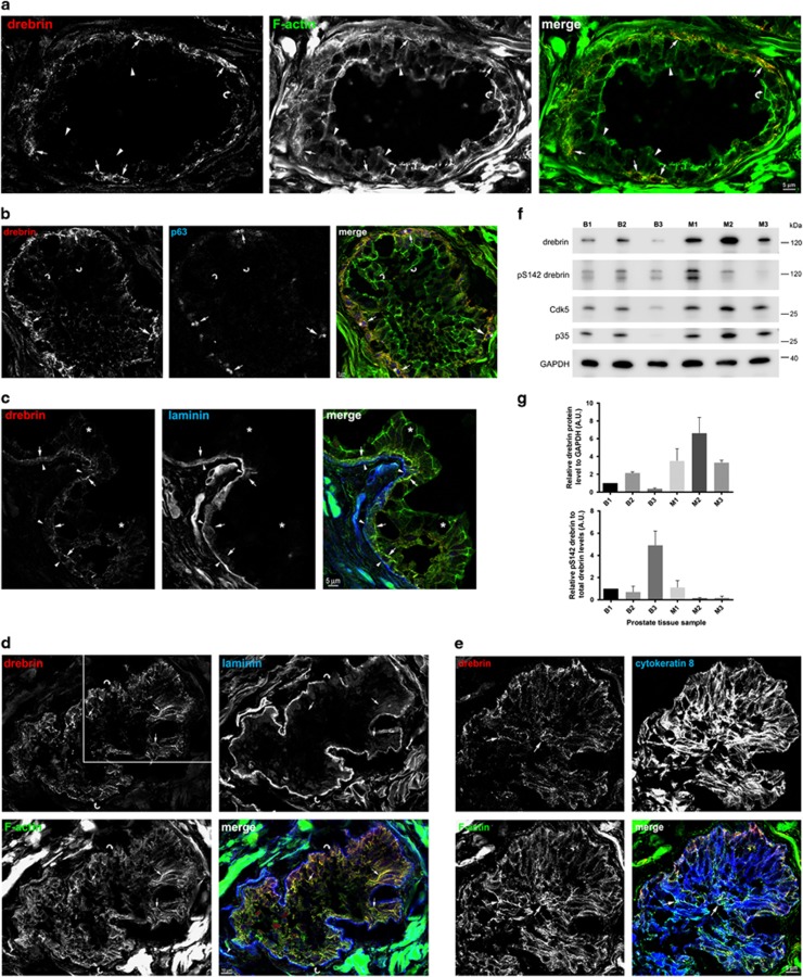
Figure 1.
Drebrin is expressed in basal epithelial cells in non-malignant human prostate and upregulated in luminal epithelial cells in human prostate cancer tissue. (a) Drebrin is expressed by a population of cells in the glandular epithelium of benign human prostate hyperplasia, where it co-localizes with F-actin. Immunofluorescence images of human prostate tissue labelled with an antibody to drebrin and phalloidin to label F-actin. Drebrin in basal cells co-localizes with F-actin (arrows). Luminal cells (arrowheads) do not contain drebrin and therefore drebrin is not associated with the F-actin in the terminal junctional web of luminal cells (curved arrow). (b) Drebrin-expressing epithelial cells also express the transcription factor protein p63, a basal cell marker, in their nucleus. Immunofluorescence images of human non-malignant prostate tissue labelled with antibodies to drebrin and p63 and with phalloidin to label F-actin. Drebrin (arrows) is expressed in cells that also express nuclear p63 (asterisks) and co-localizes with F-actin (arrows). Drebrin is not associated with the F-actin in the terminal junctional web of luminal cells (curved arrows). (c) Drebrin is expressed by cells that contact the basal lamina in the glandular epithelium of non-malignant human prostate. Immunofluorescence images of human prostate tissue labelled with antibodies to drebrin and to laminin to label the basal lamina, and with phalloidin to label F-actin. Drebrin is present in basal cells (arrows) that contact the basal lamina (arrowheads) and co-localizes with F-actin. Luminal epithelial cells do not express drebrin (asterisks). (d) Drebrin is upregulated in luminal epithelial cells in the glands of malignant human prostate tissue. Immunofluorescence images of malignant human prostate tissue labelled with antibodies to drebrin and laminin, to label the basal lamina and phalloidin to label F-actin. In luminal epithelial cells drebrin is particularly localized to baso-lateral bundles of F-actin (arrows). The glands have a disorganized architecture and there are no F-actin terminal webs visible. Notably the basal lamina appears to be deficient as indicated (curved arrows). (e) Luminal epithelial cells upregulating drebrin express cytokeratin 8, confirming their luminal phenotype. Immunofluorescence images of malignant human prostate tissue labelled with antibodies to drebrin and cytokeratin 8, and with phalloidin to label F-actin. Drebrin is expressed in cells throughout the acinus including luminal cells that also express cytokeratin 8, a marker for luminal cells (arrows). Drebrin in these cells co-localizes with F-actin. The acinus is the same one shown in d (white box area) but from an adjacent section. (f) Immunoblot of three individual cases of benign human prostate hyperplasia (B1–B3) and three individual cases of human prostate cancer (M1–M3) probed with antibodies to drebrin, pS142-drebrin, Cdk5, p35 and GAPDH, as a loading control. Drebrin is expressed in benign prostate tissue and upregulated in malignant prostate tissue. Despite upregulation of Cdk5 and p35 in malignant prostate, pS142-drebrin is variably expressed in malignant prostate. The lower band of the pS142-drebrin blot is a breakdown product of drebrin. (g) Quantification of relative drebrin and pS142-drebrin protein levels normalized to GAPDH and drebrin, respectively, from immunoblots of benign (B1–B3) and malignant (M1–M3) prostate tissue. Error bars are mean±s.e.m. from three replicate samples.

